
Edible
Requires preparation

Poisonous when raw

Set your location to see data for your region.
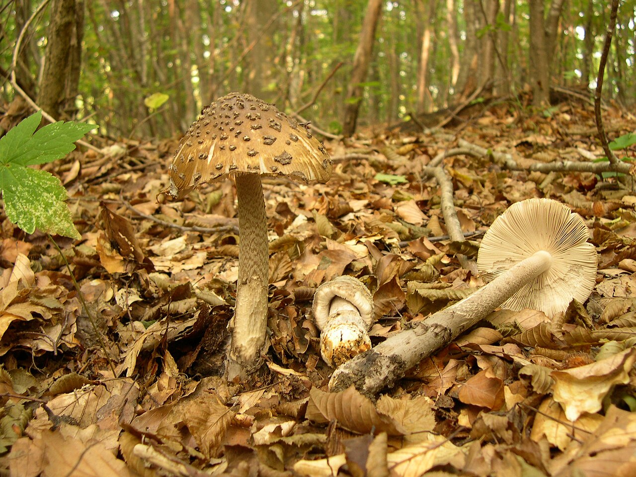
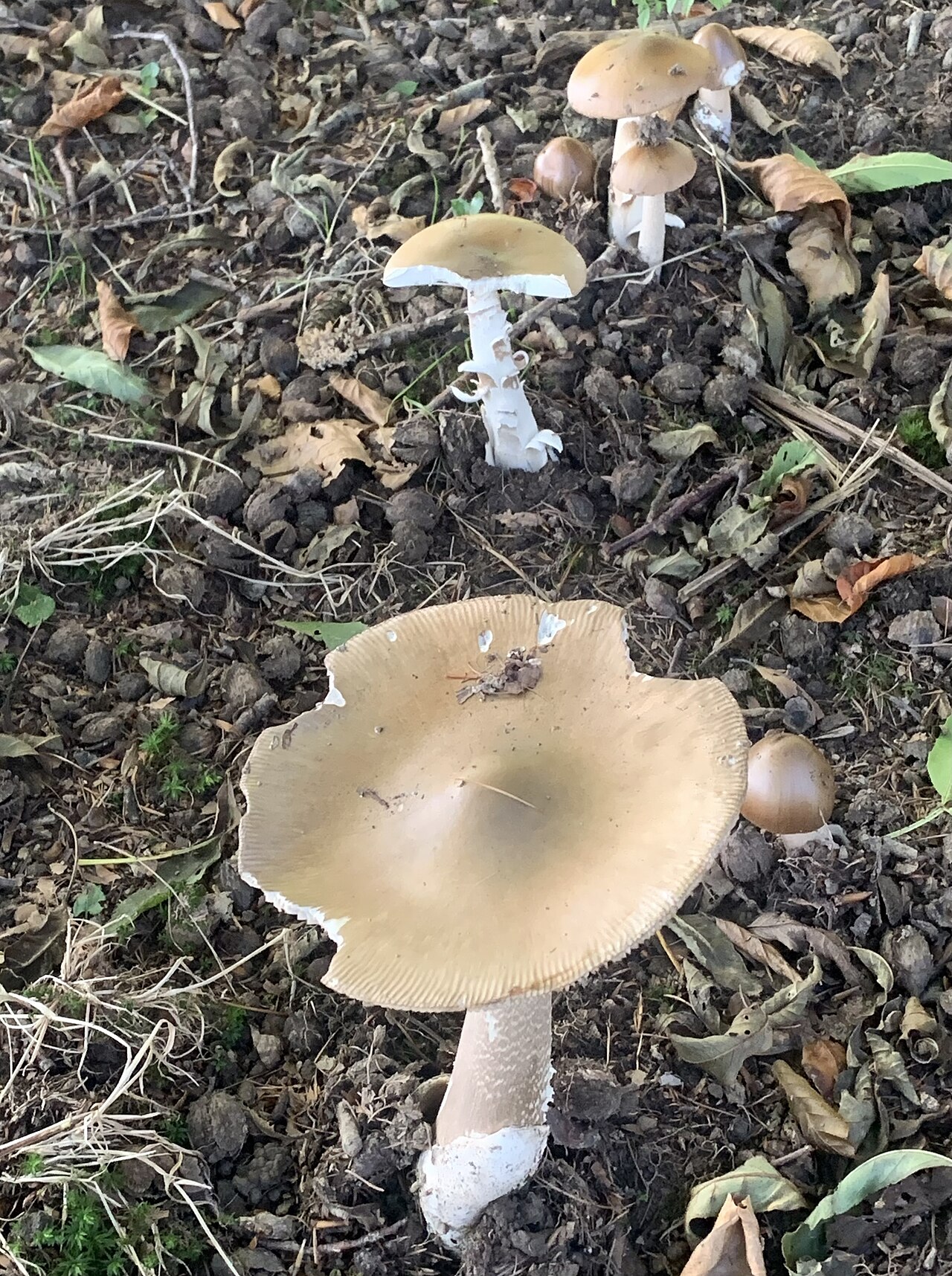
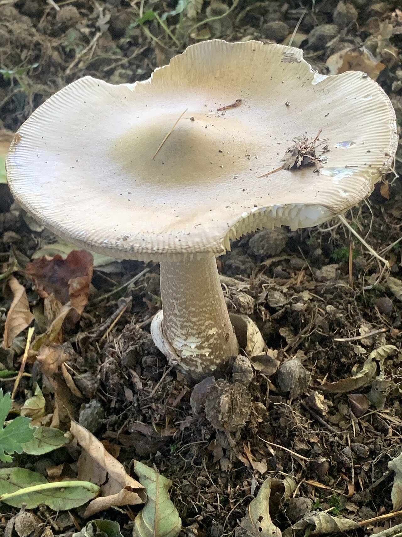
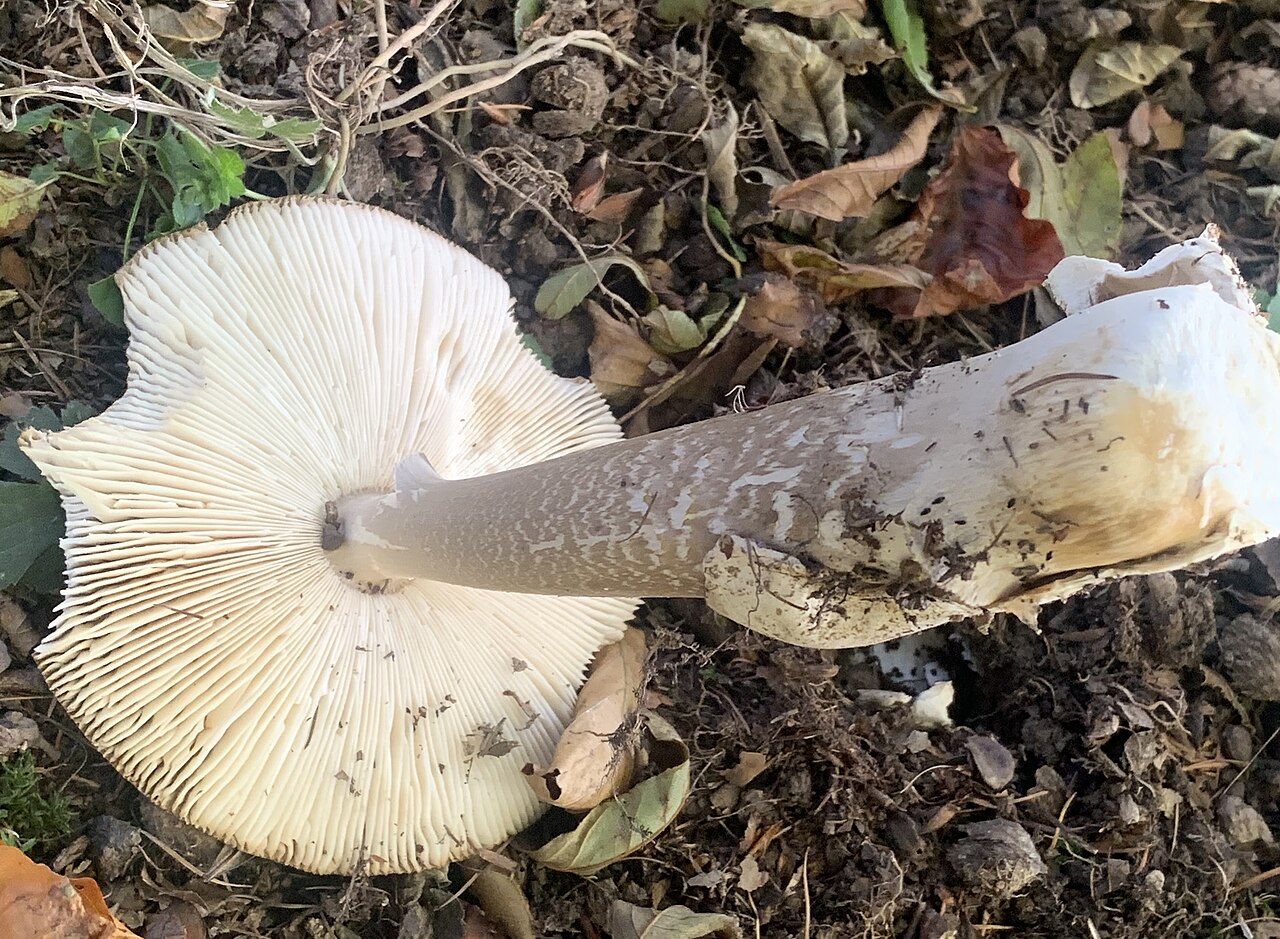
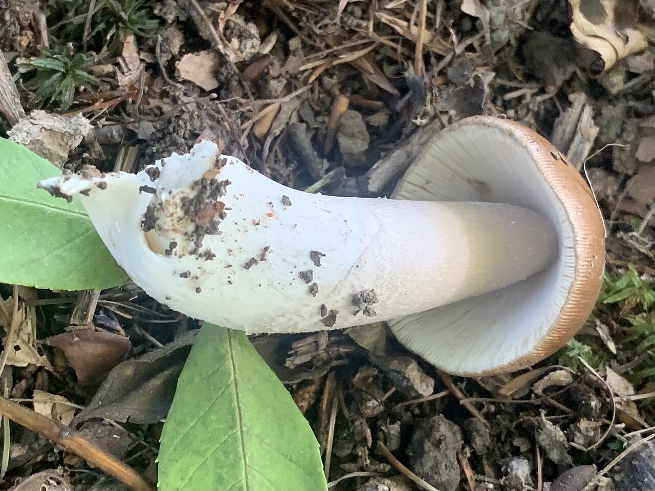
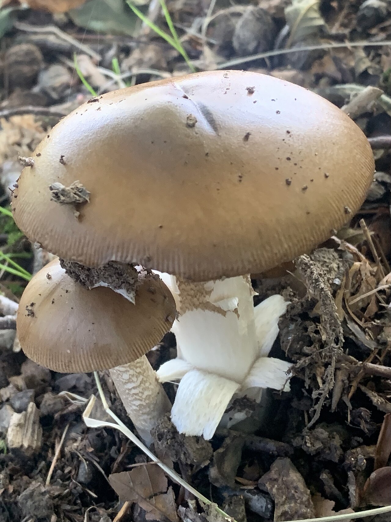
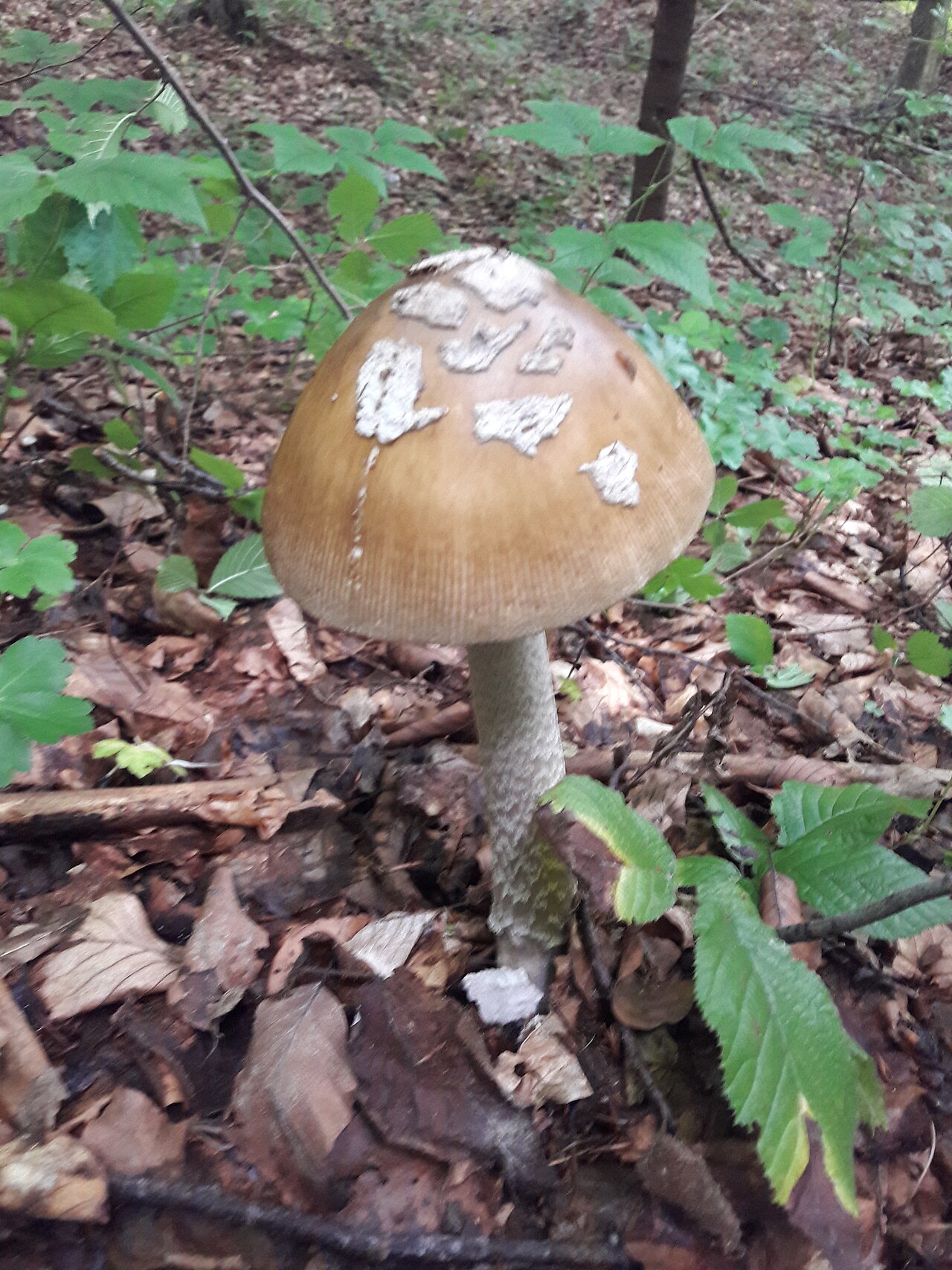
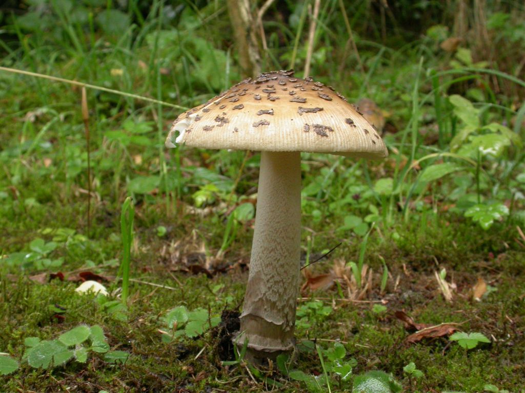
Amanita ceciliae, commonly called snakeskin grisette or Cecilia's ringless amanita, is a robust, ringless species recognizable by its large, tawny to dark yellowish-brown cap bearing charcoal-gray to brownish floccose universal veil patches. The tall, fragile stipe features distinctive zigzag concentric bands reminiscent of snakeskin, terminating in an indistinct, crumbly volva that quickly fragments into fragile zones around the stipe base rather than forming an intact bag.
Identification, habitat, and key differences.
Forager
Fruitbody structure
- Fruit body form
- Fruit body form: Cap, stipe.
Cap
- Cap shape
- Cap shape: ovoid.
Cap shape: flat.
in young fruit bodies: Cap shape: ovoid.
in mature fruit bodies: Cap shape: hemispherical.
in mature fruit bodies: Cap shape: bell-shaped.
in mature fruit bodies: Cap shape: convex.
in mature fruit bodies: Cap shape: flat.
Cap shape: convex. - Cap surface sheen
- when dry: Cap surface sheen: matt.
- Cap stickiness and sliminess
- when wet: Cap stickiness and sliminess: sticky.
- Cap colour
- Cap colour: grey-brown.
Cap colour: olive-brown.
Cap colour: reddish brown.
Cap colour: yellow-brown.
Cap colour: grayish.
Cap colour: yellow.
Cap colour: buff.
Cap colour: olive.
Cap colour: chestnut. - Margin orientation
- in old fruit bodies: Margin orientation: turned upwards.
Margin orientation: inrolled. - Cap margin surface relief
- Cap margin surface relief: grooved.
- Cap diameter
- The cap measures 50–200 mm in diameter.
- Cap margin grooving extent
- Cap margin grooving extent: 0.25 (relative to the cap radius).
Cap underside
- Hymenophore type
- Hymenophore type: gills.
Gills at stipe
- Hymenophore attachment to the stipe
- Hymenophore attachment to the stipe: attached to the stipe (gills).
Hymenophore attachment to the stipe: free from the stipe (gills).
Hymenophore attachment to the stipe: free from the stipe (gills).
- Attachment width of the hymenophore
- Attachment width of the hymenophore: narrowly attached (gills).
- General gill profile
- General gill profile: linear (gills).
- Hymenophore colour
- Hymenophore colour: grey.
Hymenophore colour: whitish.
Hymenophore colour: creamy white. - Gill branching
- Gill branching: forked.
- Macroscopic structure of the free gill edge
- Macroscopic structure of the free gill edge: crenate and floccose.
- Natural colour of the gill edge
- Natural colour of the gill edge: blackish.
- Gill spacing
- Gill spacing: closely spaced.
Gill spacing: closely spaced.
Gill spacing: crowded. - Gill thickness
- Gill thickness: thick.
- Presence of short gills
- Presence of short gills: present.
Stem and base
- Stipe shape
- Stipe shape: cylindrical.
- Stipe colour
- in the upper part of the stipe: Stipe colour: white.
in the middle part of the stipe: Stipe colour: brownish.
in the lower part of the stipe: Stipe colour: brownish.
Stipe colour: white.
Stipe colour: grey-brown. - Stipe surface covering
- Stipe surface covering: floccose.
Stipe surface covering: scaly.
in the upper part of the stipe: Stipe surface covering: powdery.
in the upper part of the stipe: Stipe surface covering: silky.
in the lower part of the stipe: Stipe surface covering: scaly. - Stem surface pattern
- Stem surface pattern: zigzag.
- Colour of stipe ornamentation
- Colour of stipe ornamentation: whitish.
- Stipe interior
- Stipe interior: solid.
Stipe interior: hollow. - Stipe consistency
- Stipe consistency: brittle.
- Stipe length
- Stipe length: 80–250 mm.
- Stipe diameter
- Stipe diameter: 10–40 mm.
- Volva
- Volva: present.
Ring
- Ring presence
- Ring presence: no ring.
Bulb
- Basal stem bulb presence
- Basal stem bulb presence: absent.
Universal veil at base
- Veil structure at the stipe base
- Veil structure at the stipe base: bands.
Veil structure at the stipe base: bands.
Veil structure at the stipe base: sac-shaped.
- Resistance of the volva and basal veil to damage
- Resistance of the volva and basal veil to damage: easily damaged (veil structure at the stem base: bands).
- Stipe flesh resistance to breaking or tearing
- Stipe flesh resistance to breaking or tearing: brittle.
- Color of volva and veil at stem base
- Color of volva and veil at stem base: white (veil structure at the stem base: volva).
Flesh
- Flesh colour
- Flesh colour: white.
- Flesh taste
- Flesh taste: sweet.
- Flesh resistance to breaking or tearing
- Flesh resistance to breaking or tearing: brittle.
- Flesh odour
- Flesh odour: not distinctive (flesh).
Flesh odour: weak.
Where and when it grows
- Habitat
- Habitat: deciduous woodland.
Habitat: coniferous woodland.
Habitat: mixed woodland.
Habitat: park.
Habitat: coniferous woodland, deciduous woodland or mixed woodland. - Substrate
- Substrate: soil.
- Fruit body growth pattern
- Fruit body growth pattern: solitary.
Fruit body growth pattern: scattered groups.
Fruit body growth pattern: in small groups. - Occurrence frequency
- Occurrence frequency: uncommon.
- Fruiting season
- Fruit bodies appear from June to October.
- Distribution area
- Distribution area: confirmed.
Edibility and culinary use
- Edibility
- Edible
- Required processing
- Requires preparation
- Raw consumption
- Poisonous when raw
Law
- Protection
- It is not subject to species protection in Poland.
- Trade
- It is approved in Poland for neither trade nor the production of mushroom products.
Possible confusions
Jerzy Opioła · CC BY-SA 3.0
Wersja JPEG o szerokości 1280 px przygotowana przez Wikimedia Commons; bez dodatkowych lokalnych zmian.
Photo source · CC BY-SA 3.0Amanita submembranacea
Key differences
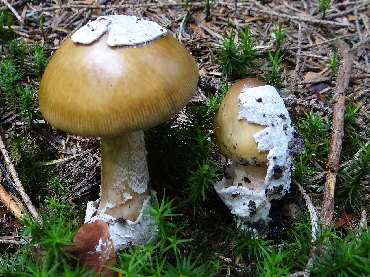
Muchomor brązowooliwkowy (Amanita submembranacea) differs in having large veil remnants on the cap and an internally greying volva, growing mainly in mountain forests on acidic soils.
Jerzy Opioła · CC BY-SA 4.0
Zmniejszono do maksymalnie 1600 pikseli i zapisano jako JPEG; zachowano proporcje, pełny kadr i barwy. Oryginalny plik źródłowy zachowano.
Photo source · CC BY-SA 4.0Amanita vaginata
Key differences
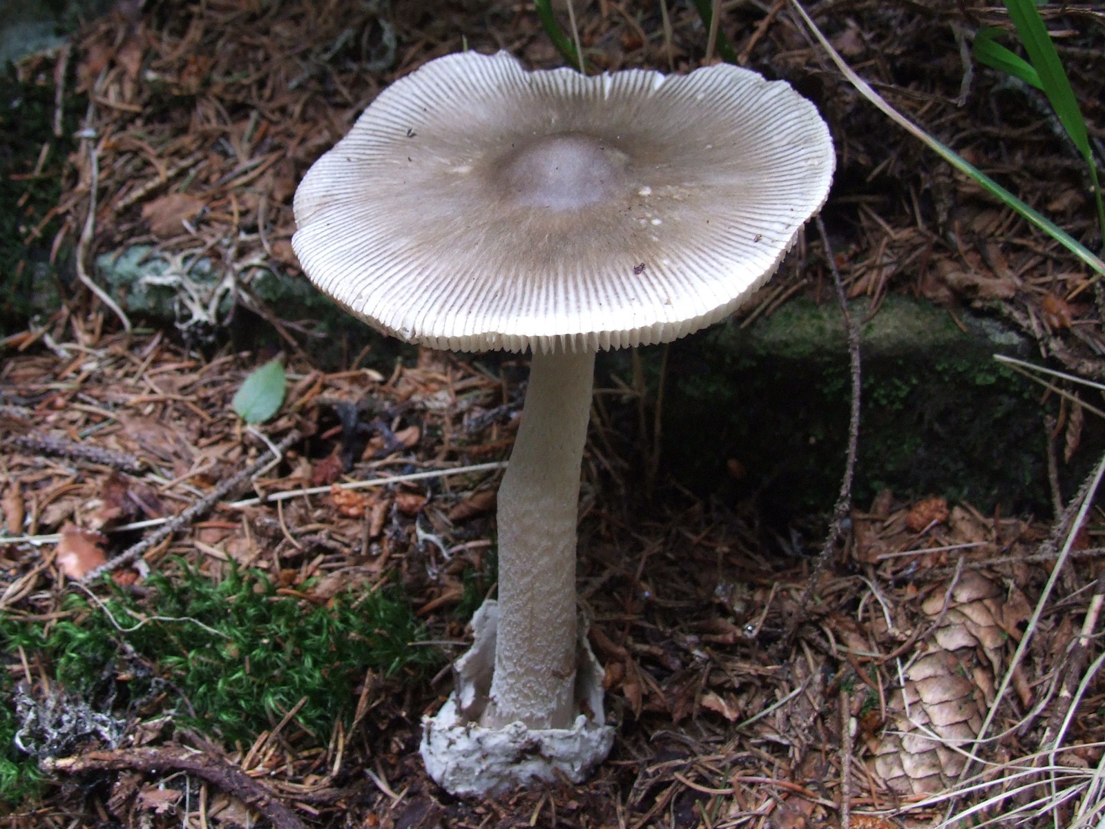
The grey amanita (Amanita vaginata) is characterized by a smooth, white stipe devoid of dark scales and a zigzag pattern.
Jerzy Opioła · CC BY-SA 4.0
Zmniejszono do maksymalnie 1600 pikseli i zapisano jako JPEG; zachowano proporcje, pełny kadr i barwy. Oryginał zachowano.
Photo source · CC BY-SA 4.0Amanita crocea
Key differences
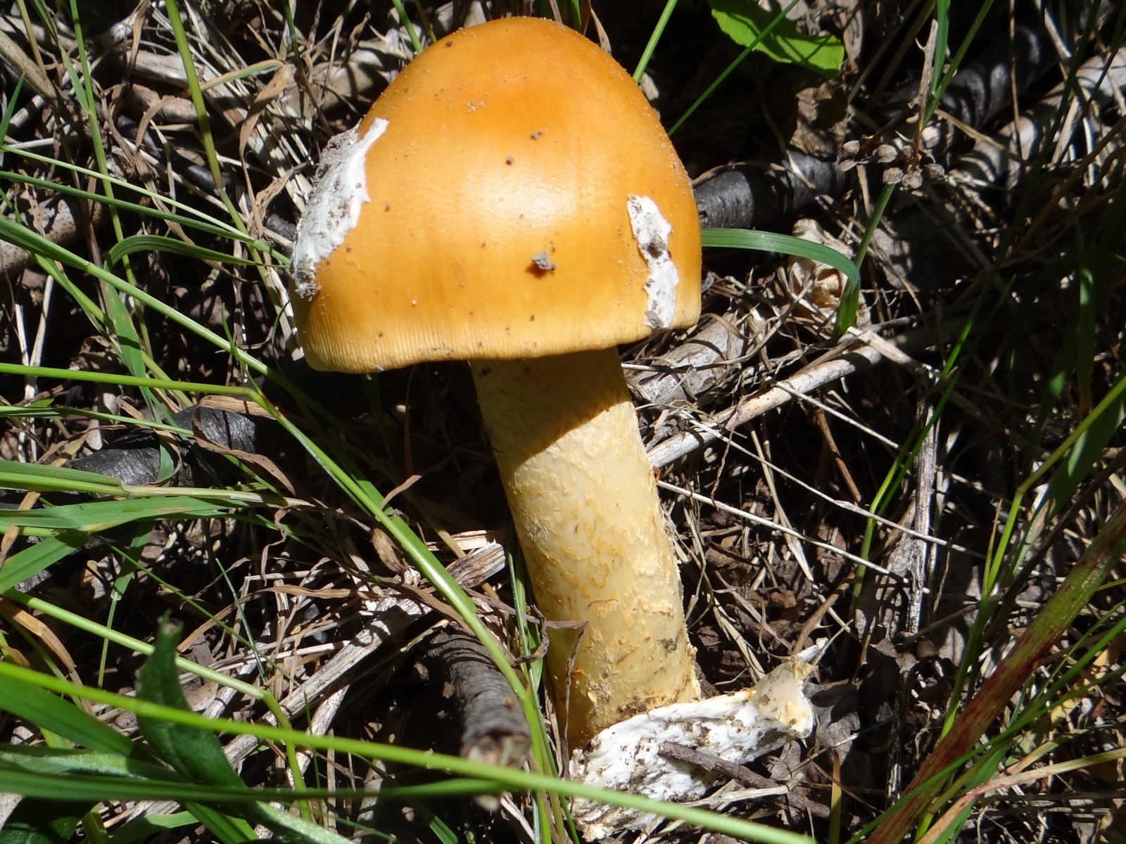
The saffron ringless amanita (Amanita crocea) has a vivid saffron to orange-yellow cap and a volva colored orange on the inside.
Amanita pantherina
Key differences
The species may be confused with the highly poisonous panther cap (Amanita pantherina). The panther cap has a distinct ring on the stipe and a bulb with concentric rims at the base, but in rain or in older specimens the ring may become worn away and fall off.
Amanita friabilis
Key differences
Amanita friabilis is significantly smaller (cap up to 6 cm), has smaller spores, and occurs exclusively under alders.
Amanita fulva
Key differences
The tawny grisette (Amanita fulva) has a cap in fox-red and buff-orange shades, a smooth stipe, and a rusting volva.
FAQ
Why is this mushroom named ceciliae?
It was named by mycologist M. J. Berkeley in honor of his wife and illustrator, Cecilia.
Can Amanita ceciliae be eaten raw?
No, consuming it raw causes severe gastrointestinal upset due to heat-labile toxins.
Does this species have a distinct ring on its stalk?
No, Amanita ceciliae lacks an annulus on its stem, typical of ringless grisettes.
What gives the stem its snakeskin appearance?
Brownish zigzag zones of floccose scales create a patterned snakeskin texture.
Which trees are the primary host partners?
It partners mainly with deciduous trees such as oak, beech, hornbeam, and chestnut.
What type of soil does it usually grow in?
It prefers rich, neutral to calcareous loam and clay soils beneath mature leaf mulch.
What is the typical fruiting season?
Fruiting takes place from mid-summer through autumn, typically July to October.
What colors does it produce when used as a textile dye?
When processed with metal mordants, it yields earthy taupe, tan, and muted gray-browns.
How does its volva differ from that of the common grisette?
Its volva is fragile and powdery, breaking into scattered rings rather than a deep sack.
Is it widely consumed in modern cooking?
No, it is generally avoided due to cooking requirements and inconsistent tolerance.
Enthusiast
Names and history
- Obowiązująca nazwa krajowa
- Muchomor złotawy
- Obowiązująca nazwa taksonomiczna
- Amanita ceciliae (Berk. & Broome) Bas
- Inne nazwy krajowe
- Snakeskin Grisette, Cecilia’s Ringless Amanita, Strangulated Amanita
- Pozostałe nazwy
- muchomor pochwiasty forma łuskowata, muchomor wężowoskóry, gryzetka złotawa, mglejarka wężowoskóra, mglejarka złotawa, podsadka złotawa
Home observations
Spore print
- Required materials
- fresh, mature cap, sheet of paper, glass or small bowl, water
- Time
- 2–24 h
- Procedure
- Place the cap underside down on the paper.
- Add a drop of water on top and cover.
- After 2–24 hours, lift the cap and inspect the deposit.
What to look for
- Spore print colour
- Spore print colour: white.
Under the microscope
- Required materials
- light microscope, microscope slide and cover slip, needle, water, spores from print
- Procedure
- Transfer a small amount of the spore print onto the slide using a needle.
- Add a drop of water, cover with a cover slip, and examine the spores.
What to look for
- Spore shape
- Spore shape: globose. Spore shape: globose. Spore shape: subglobose. Spore shape: globose. Spore shape: globose (nearly). Spore shape: globose. Spore shape: globose (nearly).
- Spore surface
- Spore surface: smooth. Spore surface: smooth.
- Spore dimensions
- Length 9.5–25 µm, width 8.6–25 µm.
Compare dimensions after calibrating the microscope scale.
Ecology and distribution
- Soil calcareousness
- Soil calcareousness: calcareous.
- Broadleaf trees in the vicinity
- Broadleaf trees in the vicinity: beech.
Broadleaf trees in the vicinity: hornbeam.
Broadleaf trees in the vicinity: oak. - Coniferous trees in the vicinity
- Coniferous trees in the vicinity: fir.
- Mode of nutrition
- Mode of nutrition: Mycorrhizal.
Non-culinary uses
Amanita ceciliae has no widespread industrial or medical application, but it has recognized non-culinary utility in historical botanical art, dye craft, and biological education. The distinctive contrast between its brownish pileus and dark universal veil fragments made it a frequent subject of Victorian scientific watercolorists and anatomical modelers studying fungal veil differentiation.
In fiber craft experiments, amateur dyers have tested mature caps with alum, copper, and iron mordants, yielding soft, earthy taupe, gray-brown, and muddy mustard tones on natural raw fleece and linen yarn. Beyond craft uses, dried specimens are widely maintained in national fungaria and university collections as morphotaxonomic references, demonstrating the mechanical transition between membranous volvas and friable, pulverulent veil remnants in basal Amanita evolutionary biology.
History and culture
Amanita ceciliae holds a unique and celebrated place in nineteenth-century British botanical history. Described in 1854 by the eminent British mycologist Reverend Miles Joseph Berkeley and Christopher Edmund Broome, it was specifically named in honor of Cecilia Berkeley (née Campbell), Berkeley's wife. She was an accomplished botanical artist and collaborator who prepared detailed illustrations, collected specimens, and assisted in his extensive mycological correspondence.
In historical literature, the mushroom was sometimes referred to under the dramatic moniker 'Amanita strangulata' in reference to the constricted appearance of its fragmented volva. In British and Victorian natural history circles, the species represented the quiet contributions of women to nineteenth-century science, immortalizing Cecilia Berkeley in official biological nomenclature. In countryside folklore, the snakeskin patterning on the stipe gave rise to superstitious associations with serpents and wood spirits, prompting rural travelers to view the imposing, spotted caps with wary curiosity.
Edibility and safety
Amanita ceciliae is considered conditionally edible upon thorough cooking, as the raw tissues contain thermolabile toxins that cause painful stomach cramps, nausea, and gastrointestinal irritation. High heat applied over an extended cooking period is mandatory to break down these harmful components and render the flesh digestible.
Even after proper heating, eating this species is widely discouraged by modern foraging societies due to the fragility of its flesh, its mediocre culinary quality, and significant digestive variability among consumers. In traditional rural cuisine in parts of Southern and Eastern Europe, it was historically collected and cooked after parboiling, valued for a sweet, earthy, though somewhat watery taste. Conversely, in the United Kingdom and North America, it is classified almost universally as non-recommended or inedible due to safety margins and negligible table worth.
Habitat and occurrence
Amanita ceciliae is widely distributed throughout temperate regions of Europe and temperate parts of Asia, whereas broad reports asserting a widespread North American distribution reflect misapplied records of other taxa rather than confirmed widespread presence. It thrives as an ectomycorrhizal mutualist within mature deciduous and mixed woodlands, establishing partnerships primarily with oak (Quercus), beech (Fagus sylvatica), hornbeam (Carpinus betulus), and sweet chestnut (Castanea sativa). Occasionally, it is recorded under coniferous associates such as silver fir (Abies alba) and pine in montane mixed stands.
The species favors warm-temperate microclimates, calcareous to neutral loam soils, and clay-rich substrates covered with rich leaf mulch, frequently fruiting along dappled glades, shaded banks, and ancient woodland paths. Its fruiting period extends from late June or July through October, with peak emergence during warm, humid late-summer periods following heavy rainfall. In Mediterranean regions, fruiting may persist into November, while northern European populations finish production earlier in autumn as temperatures decline.
Researcher
Edibility and culinary use
Ustalenia źródłowe: jadalność, spożycie i wykorzystanie
- Edibility
- Edible — źródło oznaczenia
Karel Tejkal/Česká mykologická společnost, Amanita ceciliae — atlas i bibliografia · Culinary use, author's assessment, without clinical safety study
- Raw consumption
- Poisonous
Materiał: surowy
SFMU, materiał dydaktyczny Champignons pod kierunkiem V.Danela, rewizja grudzień2019 · Amanita inaurata* list: toxic raw or undercooked; educational source, not species toxin analysis
- Edibility
- Poisonous
Materiał: po niedostatecznej obróbce
SFMU, materiał dydaktyczny Champignons pod kierunkiem V.Danela, rewizja grudzień2019 · Amanita inaurata* list: toxic raw or undercooked; educational source, not species toxin analysis
- Required processing
- Requires preparation
Wynika z potwierdzonej toksyczności materiału surowego.
SFMU, materiał dydaktyczny Champignons pod kierunkiem V.Danela, rewizja grudzień2019 · Lista Amanita inaurata*: toksyczne surowe lub niedogotowane; źródło dydaktyczne, nie analiza toksyny gatunku
Scientific names and taxonomy
- Current scientific name
Amanita ceciliae (Berk. & Broome) Bas
Scientific names and taxonomy
Amanita ceciliae (Berk. & Broome) Bas belongs to the family Amanitaceae (Amanitaceae) and section Vaginatae. It was described in 1854 by Berkeley and Broome under the basionym Agaricus ceciliae, and Cornelis Bas transferred it to the genus Amanita in the 80s of the 20th century.
Scientific names and taxonomy
In earlier mycological publications, this species appeared, among others, under the names Amanita inaurata, Amanitopsis inaurata, and Amanitopsis ceciliae. The name Amanita strangulata was, in turn, used by many authors in a broad, ambiguous sense (sensu auct. mult.).
Historical scope of names and species concepts
In English-language literature, the name Snakeskin Grisette is used, and in German, Riesen-Scheidenstreifling.
Taxon scope and identification limitations
Taxonomic studies have shown that some North American mushrooms previously described as A. ceciliae or A. inaurata belong to distinct species, such as Amanita rhacopus and Amanita variicolor. The modern concept of A. ceciliae refers to European populations.
Taxon scope and identification limitations
In phylogenetic studies of section Vaginatae, the position of the European taxon A. ceciliae (represented, among others, by collections from Hungary) was confirmed using sequence analysis of four loci: ITS, LSU, RPB1, and TEF1-α.
- Zakres ujęcia taksonomicznego
Profil obejmuje doniesienia z niezależnych europejskich źródeł pod nazwą Amanita ceciliae, w tym osobno udokumentowany węgierski okaz dowodowy DB-2018-07-08-5/AD22. Nie stanowi ponownego oznaczenia wszystkich okazów opisanych w atlasach ani nie utożsamia z materiałem europejskim meksykańskich ujęć inaurata/ceciliae, ujęcia Kaqchikel aff. ceciliae, A. sororcula lub innych ujęć pozaeuropejskich.
[13] [10] [14] [3]
Ecology and distribution
Laboratory diagnostics
Dane ustrukturyzowane
Basidia
- Basidium sterigma length
- Basidium sterigma length: up to 8.4 µm. [34]
- Spores per basidium
- The number of spores per basidium is 4. [36]
The number of spores per basidium is 2. [36]
The number of spores per basidium is 2. [34]
The number of spores per basidium is 4. [34] - Basidium shape
- Basidium shape: clavate. [36]
- Sterigma width
- Sterigma width: up to 3.1 µm. [34]
- Clamp connections
- Clamp connections: not observed. [36]
Other laboratory characteristics
- Gill edge fertility
- Gill edge fertility: sterile. [36]
- Subhymenial zone measurement
- Subhymenial zone measurement: 15–35 µm (wst-near measurement). [34]
Subhymenial zone measurement: 50–70 µm (wst-far measurement). [34] - Arrangement of universal veil cells
- Arrangement of universal veil cells: terminal (on the cap). [34]
Arrangement of universal veil cells: in short rows (on the cap). [34]
Pileipellis
- Optical appearance of pileipellis hyphae
- Optical appearance of pileipellis hyphae: hyaline (upper layer of the cap cuticle). [36]
- Arrangement of pileipellis hyphae
- Arrangement of pileipellis hyphae: nearly radial. [34]
- Shape of pileipellis elements
- Shape of pileipellis elements: cylindrical. [36]
Shape of pileipellis elements: cylindrical. [36] - Width of pileipellis elements
- Width of pileipellis elements: 1.1–7.7 µm. [34]
Width of pileipellis elements: 0.8–3 µm. [36]
Width of pileipellis elements: 2.5–4 µm. [36] - Branching of the pileipellis hypha
- Branching of the pileipellis hypha: branched. [34]
- Embedding of cuticle hyphae in a gelatinous substance
- Embedding of cuticle hyphae in a gelatinous substance: present. [36]
Embedding of cuticle hyphae in a gelatinous substance: present (at the surface only, in the surface layer of the pileipellis). [34]
Embedding of cuticle hyphae in a gelatinous substance: no gelatinization (in the lower pileipellis layer). [34]
Embedding of cuticle hyphae in a gelatinous substance: present (at the surface only, in the surface layer of the pileipellis, in a mature fruiting body). [34] - Color of cuticle pigment
- Color of cuticle pigment: brown. [34]
Reactions with reagents
Spores
- Mean spore length-to-width ratio
- Mean spore length-to-width ratio: 1.04 (mean). [32]
- Profile of the adaxial side of the basidiospore
- Profile of the adaxial side of the basidiospore: flattened. [34]
- Presence of the basidiospore hilar appendix
- Presence of the basidiospore hilar appendix: present. [34]
- Position of the basidiospore hilar appendix
- Position of the basidiospore hilar appendix: sublateral. [34]
- Basidiospore hilar appendix shape
- Basidiospore hilar appendix shape: cylindrical. [34]
- Spore colour under the microscope
- Spore colour under the microscope: hyaline. [36]
Spore colour under the microscope: hyaline. [34] - Spore length
- Spore length: 9.7–15.4 µm. [36]
Spore length: 10.5–14 µm. [10]
Spore length: 9.5–25 µm. [34] - Presence of lipid bodies
- Presence of lipid bodies: present. [36]
- Spore length-to-width ratio
- Spore length-to-width ratio: 0.94–1.14. [36]
Spore length-to-width ratio: 1–1.36. [34] - Spore shape
- Spore shape: globose. [10]
Spore shape: globose. [9]
Spore shape: subglobose. [9]
Spore shape: globose. [36]
Spore shape: globose (nearly). [36]
Spore shape: globose. [34]
Spore shape: globose (nearly). [34] - Spore volume
- Spore volume: 410–2030 µm³. [36]
- Spore wall thickness
- Spore wall thickness: thin-walled. [34]
Hymenophoral trama
- Hymenophoral trama type
- Hymenophoral trama type: bilateral. [34]
- Width of trama hyphae
- Width of trama hyphae: 2.8–5.9 µm. [34]
- Length of inflated hymenophoral trama cell
- Length of inflated hymenophoral trama cell: up to 74 µm. [34]
- Width of inflated hymenophoral trama cell
- Width of inflated hymenophoral trama cell: up to 16.7 µm. [34]
Material and research methods
Description of characteristics — source and scope
Material: Atlas description of flesh by K.Tejkal/ČMS; odor nevýrazný.
Readout scope: Documented observation.
Limitations: Author description without specimen number; indistinct odor was not equated with chemical absence of odor substances.
Microscopic data — source and limitations
Material: Mean Q of spores from the collection of 4813,100 measurements by author M. Zugna; the same material as a separate microscopic examination, not a new experiment.
Readout scope: Documented observation.
Limitations: Mean reported directly by the author; independent length and width boundaries were not separated.
Study of composition and activity — Akata et al. (2012)
Material: Fruit bodies from Anatolia, identified as Amanita ceciliae, AKATA 3037,850 m; methanol extract of dried material. DPPH assay, 517 nm, 30 min in the dark; three replicates.
Readout scope: Documented observation.
Limitations: In vitro result, not a confirmed treatment or safe food dose. No molecular verification of identification in this work; non-Polish material. The abstract contains a contradictory assignment of 96,16% at 4,51 mg/ml; the directly read row gives 95,35 ± 0,10% at 4,65 mg/ml.
Archiwalny opis siedliska
Krajowe zbiory udokumentowano m.in. w Beskidzie Niskim (okolice Chyrowej, Myscowej, Woli Niżnej i Tylawy) w drzewostanach jodłowo-bukowych z domieszką grabu, leszczyny i wiązu.
[10]Obecność gatunku potwierdzają również historyczne zbiory zielnikowe z lasów grądowych Puszczy Niepołomickiej.
[11]Archiwalny opis siedliska
W Europie Środkowej owocnikowanie trwa od czerwca do października, a grzyb występuje miejscami obficie w ciepłolubnych lasach dębowych i bukowych.
[6]- Results as recorded in the sources — Spores per basidium
- Liczba zarodników na jednej podstawce wynosi 2. [36] [34]
Liczba zarodników na jednej podstawce wynosi 4. [36] [34] - Results as recorded in the sources — Width of the universal veil hypha
- Strzępka osłony całkowitej może mieć szerokość od 1,1 do 11,7 mikrometra. [34]
Strzępka osłony całkowitej może mieć szerokość od 2,5 do 4,5 mikrometra. [36]
Metody i zapisy źródłowe — Zbiór 4813, Prato Rotondo koło Teramo, Włochy, 19.08.2012, leg. Bruno de Ruvo, det. Marino Zugna. Suchy materiał rehydratowano wodą lub KOH 5%; preparaty w wodzie albo amoniakalnej czerwieni Kongo 6%. Nowy wynik dotyczy wyłącznie strzępek zewnętrznego pokrycia opisanego jako ixocutis, nie pigmentowanej warstwy cutis.
Wygląd optyczny strzępek skórki — zapis źródłowy: „Rivestimento pileico ... ixocutis ... ife cilindriche ... ialine; tylko strzępki tego pokrycia.” [36]
Metody i zapisy źródłowe — Wyłącznie zbiór E. W. Brown, Middleton, Dyfed, Walia, 20.09.1994, RET270-1. Cała skórka ma 155–220 µm; opis źródłowy oddziela powierzchnię, suprapellis i subpellis. Grubości poszczególnych warstw nie utożsamiono z grubością całości.
Układ strzępek skórki — zapis źródłowy: „In E. W. Brown collection: filamentous undifferentiated hyphae subradially arranged.” [34]
Osadzenie strzępek skórki w substancji żelowej — zapis źródłowy: „In E. W. Brown collection: completely gelatinized only at surface; zakres przestrzenny ograniczony do powierzchni.” [34]
Osadzenie strzępek skórki w substancji żelowej — zapis źródłowy: „In E. W. Brown collection: subpellis ... ungelatinized; dokładny wynik ujemny wyłącznie tej warstwy.” [34]
Grubość skórki kapelusza — zapis źródłowy: „In E. W. Brown collection: pileipellis 155–220 µm thick; cała skórka.” [34]
Metody i zapisy źródłowe — Rewizja europejskich eksykatów R.E.Tullossa: 100pomiarów z5owocników i4zbiorów. Lista materiału obejmuje Perreau475(Francja),BrownRET270-1(Walia),KibbyRET270-2(Anglia),Gminder96/141=RET305-4(Niemcy) i historyczny materiał Berkeley. Wymiary zbiorcze nie zostały przypisane jednemu z tych okazów.
Długość sterygmy podstawki — zapis źródłowy: „Do8,4µm długości; szerokość3,1µm oczekuje osobnego pola.” [34]
Liczba zarodników na jednej podstawce — zapis źródłowy: „Podstawki4sterygmowe i rzadko2sterygmowe.” [34]
Profil adaksjalnej strony bazydiospory — zapis źródłowy: „Spłaszczone od strony adaksjalnej” [34]
Położenie wyrostka wnęki bazydiospory — zapis źródłowy: „Podboczny” [34]
Obecność wyrostka wnęki bazydiospory — zapis źródłowy: „Wyrostek opisany wprost” [34]
Kształt wyrostka wnęki bazydiospory — zapis źródłowy: „Cylindryczny” [34]
Długość podstawki; Szerokość podstawki; Długość zarodnika; Iloraz długości i szerokości zarodnika; Szerokość zarodnika — zapis źródłowy: „Pełne potwierdzone granice, z zachowaniem zakresów centralnych i zbiorczych średnich w źródle.” [34]
Typ tramy hymenoforu — zapis źródłowy: „Bilateralna” [34]
Szerokość strzępek tramy — zapis źródłowy: „Nitkowate strzępki2,8–5,9µm; nie zakres naczyniowych2,8–23µm.” [34]
Długość rozdętej komórki tramy hymenoforu — zapis źródłowy: „Do74µm; nie przepisano tego wymiaru strzępkom naczyniowym.” [34]
Szerokość rozdętej komórki tramy hymenoforu — zapis źródłowy: „Do16,7µm, własna szerokość opisanych komórek.” [34]
Barwa zarodnika w mikroskopie — zapis źródłowy: „Bezbarwne, hialinowe, bez określonego medium.” [34]
Kształt zarodnika — zapis źródłowy: „Kuliste do niemal kulistych; określenie przy własnym kształcie.” [34]
Powierzchnia zarodnika — zapis źródłowy: „Gładkie” [34]
Grubość ściany zarodnika — zapis źródłowy: „Cienkościenne” [34]
Szerokość strzępki osłony całkowitej — zapis źródłowy: „Nitkowate strzępki osłony” [34]
Metody i zapisy źródłowe — Włochy,Teramo,PratoRotondo,19.08.2012,1460m,las bukowy Fagus sylvatica,zbiór4813; leg.B.deRuvo,det.M.Zugna. Suchy materiał rehydratowano5%KOH lub wodą; preparaty w wodzie lub6%amoniakalnej czerwieniKongo. Mycométre,100dojrzałych zarodników z niehymenialnych części w projekcji bocznej; pozostałe typy komórek po co najmniej20pomiarów.
Liczba zarodników na jednej podstawce — zapis źródłowy: „Zwykle4zarodnikowe, rzadko2zarodnikowe; określenie dotyczy wyłącznie własnej liczby.” [36]
Długość podstawki; Szerokość podstawki; Długość zarodnika; Iloraz długości i szerokości zarodnika; Objętość zarodnika; Szerokość zarodnika — zapis źródłowy: „Pełne źródłowe min–max, wraz z granicami w nawiasach; zakresy centralne i średnie zachowano w odczycie oryginału.” [36]
Kształt podstawki — zapis źródłowy: „Maczugowate; bardzo wydłużone jako odrębna potrzeba modelu.” [36]
Płodność ostrza blaszek — zapis źródłowy: „Ostrze blaszki sterylne; nie brzeg porów.” [36]
Kształt elementów skórki kapelusza — zapis źródłowy: „Strzępki cylindryczne” [36]
Szerokość elementów skórki — zapis źródłowy: „Odrębny zakres własnej warstwy” [36]
Osadzenie strzępek skórki w substancji żelowej — zapis źródłowy: „Ixocutis, osadzenie w zżelatynizowanej warstwie; bez zgadywania stopnia.” [36]
Organizacja skórki kapelusza — zapis źródłowy: „Ixocutis: organizacja cutis, zżelatynizowanie nie jest osobnym łącznym kodem.” [36]
Organizacja skórki kapelusza — zapis źródłowy: „Cutis” [36]
Barwa zarodnika w mikroskopie — zapis źródłowy: „Hialinowe; medium tej ogólnej oceny nie przypisano odrębnie.” [36]
Obecność ciał lipidowych — zapis źródłowy: „Autor rozpoznaje centralną kroplę olejową” [36]
Kształt zarodnika — zapis źródłowy: „Kuliste do niemal kulistych; określenie przy własnym kształcie.” [36]
Kształt komórki osłony całkowitej — zapis źródłowy: „Sferocyty; średnicy17–40µm nie przepisano jako dwóch osi.” [36]
Szerokość strzępki osłony całkowitej — zapis źródłowy: „Średnica nitkowatych strzępek” [36]
Metody i zapisy źródłowe — Rewizja europejskiego materiału R. E. Tullossa. Pomiary subhymenium i sterygm oraz opis osłony są wynikami zbiorczego opisu źródła; nie przypisano ich samowolnie jednemu okazowi. Wskazane w źródle N=100 dotyczy zarodników, nie tych nowych pomiarów.
Szerokość sterygmy — zapis źródłowy: „Technical, basidia: sterigmata up to 8.4 × 3.1 µm; wyłącznie szerokość, brak dolnej granicy.” [34]
Pomiar strefy podhymenialnej — zapis źródłowy: „Technical, subhymenium: wst-near = 15–35 µm.” [34]
Pomiar strefy podhymenialnej — zapis źródłowy: „Technical, subhymenium: wst-far = 50–70 µm; inna miara niż wst-near.” [34]
Układ komórek osłony całkowitej — zapis źródłowy: „Technical, universal veil on pileus: terminal singly and in chains of up to 3 cells; położenie końcowe komórek.” [34]
Układ komórek osłony całkowitej — zapis źródłowy: „Technical, universal veil on pileus: terminal singly and in chains of up to 3 cells; krótkie szeregi. Liczba do 3 pozostaje w dokumentacji do osobnego rozszerzenia.” [34]
Kształt komórki osłony całkowitej — zapis źródłowy: „Technical, universal veil on pileus: globose to subglobose to pyriform; zapis tylko jawnego podstawowego kształtu globose.” [34]
Kształt komórki osłony całkowitej — zapis źródłowy: „Technical, universal veil on pileus: ellipsoid to subclavate; zapis wyłącznie jawnego ellipsoid, bez utraty kwalifikatora przez dopisanie clavate.” [34]
Metody i zapisy źródłowe — Wyłącznie zbiór E.W.Brown,Middleton,Dyfed,Walia,20.09.1994,RET270-1. Opis skórki odróżnia powierzchnię,częściowo zżelatynizowaną suprapellis i niezżelatynizowaną subpellis z brązowym pigmentem wewnątrz komórek.
Szerokość elementów skórki — zapis źródłowy: „Zwykłe strzępki1,1–7,7µm” [34]
Rozgałęzienie strzępki skórki — zapis źródłowy: „Rozgałęzione” [34]
Barwa pigmentu skórki — zapis źródłowy: „Brązowy pigment subpellis; położenie wewnątrzkomórkowe zapisane przy dokładnym celu.” [34]
Metody i zapisy źródłowe — Wyłącznie Perreau 475. Autor podaje w pełni rozwinięty okaz pozostawiony na publicznym pokazie przed dokładnym wysuszeniem. Skórka 65–95 µm i pełne zżelatynizowanie tylko powierzchni. Nie połączono tego okazu z kolekcją E. W. Browna.
Osadzenie strzępek skórki w substancji żelowej — zapis źródłowy: „In Perreau 475: completely gelatinized only at surface; dokładny inny okaz.” [34]
Grubość skórki kapelusza — zapis źródłowy: „In Perreau 475 (fully expanded specimen left on public display before thorough drying): pileipellis 65–95 µm thick.” [34]
Metody i zapisy źródłowe — Marek Snowarski/GRej, Amanita ceciliae
Kształt zarodnika — zapis źródłowy: „Kuliste i przejrzyste w opisie mikroskopowym; opis barwy zapisano też osobno od wysypu.” [10]
Powierzchnia zarodnika — zapis źródłowy: „Gładka.” [10]
Szerokość zarodnika — zapis źródłowy: „GRej: pełna para10,5–14×10–14 µm; druga oś10–14 µm.” [10]
Metody i zapisy źródłowe — Pat O’Reilly/FirstNature, Snakeskin Grisette
Species names and boundaries
Polish name: Muchomor złotawy is the name used in Wojewoda's Polish checklist 2003. The author refers to Borowska and Skirgiełło in Garnweidner 1993; an earlier edition was not examined. The checklist also retains Domański's historical name 1955 a. Six names from the Ekologia.pl atlas were recorded as alternative with atlas provenance. This does not establish them as official names nor does it confirm old unresolved record labels.
Original publications: Berkeley and Broome described Agaricus ceciliae in 1854, entry 663, pp. 396–397. Bas in 1984, p. 192, after examining type material at Kew, created the combination Amanita ceciliae. The year 1984 was read in the publication itself; earlier discrepancy 1983/1984 remains in history. Bas separated incorrect later uses of the name strangulatus from Fries's original description 1838, concerning a different, unresolved species. True Agaricus strangulatus was not added as a simple synonym.
Taxon scope and extra-European material: The study concerns the European concept of Amanita ceciliae, including the separately documented Hungarian specimen DB-2018-07-08-5/AD22. This does not imply redetermination of all atlas specimens nor equating Mexican, Guatemalan aff. ceciliae, or A. sororcula material with European material. Lambert and co-authors segregated A. rhacopus and A. variicolor from former North American determinations based on morphology, types, and ITS. The restriction also applies to transferring photos, poison reports, and consumption reports.
Polish occurrence and fruiting
Occurrence and months: Wojewoda lists Polish localities including the vicinity of Elbląg, Wielkopolski National Park, Kampinos, Głogów, Dębowiec, Annopol, Babia Góra, Pieniny, and Polesie, as well as the period June–October. This is a literature review, not a new monitoring of all of Poland. GRej contains further, explicitly identified Polish observations. Months were entered from the Polish checklist; individual collection dates were not converted into season boundaries.
Habitat and caution in interpretation: The Polish checklist mentions soil in deciduous, coniferous, and mixed forests, also under spruce. Snowarski describes a preference for calcareous soils and infrequent records. Presence next to a tree was not turned into molecular proof of a mycorrhizal association. The historical category of regional threat is not current national legal protection. Foreign months or red list statuses do not replace Polish data.
Examination of Italian collection 4813
Material and method: The collection from 19.08.2012 comes from a beech forest near PratoRotondo, Teramo, 1460 m; collected by Bruno de Ruvo, determined by Marino Zugna. The macro description was prepared from photographs. Dry material was rehydrated with water or 5%KOH; mounts were examined in water or 6%ammoniacal Congo red. Melzer was used to evaluate amyloid response. 100 mature spores from non-hymenial parts were measured in side view; other cell types at least 20 times each. A single medium was not assigned to all measurements.
Results: The full spore ranges are 9,7–15,4×8,9–15,8 µm, Q0,94–1,14, mean Q 1,04, volume 410–2030 µm³. They are globose to subglobose, hyaline, inamyloid, with one large central oil drop. Basidia measure 38,63–68,53×10,39–16,33 µm, usually 4, and rarely 2 spores. Clamps were not observed in the examined tissues. Central ranges, means, and source caveats were retained as in the original.
Limitations: These are the author's own observations shared by the collector on a blog; the earlier forumAMB version was not read. The specimen is not a Polish measurement. The two layers of the pellis have separate hyphal width ranges; they were not combined into a single unconditional range. The absence of observations of marginal cells is not proof of the absence of all cystidia.
Revision of European exsiccata
Material and result: Tulloss reports 100 measurements from 5 basidiocarps and 4 collections. Full spore limits are 9,5–25×8,6–25 µm, with narrower central ranges 10,3–14,9×9,5–14,3 µm; the source explicitly mentions occasional giant spores. Basidia 65–89×13,8–19,1 µm are 4 sterigmate or rarely 2 sterigmate. The medium was not specified for each measurement. Bas's notes on the type were not turned into an independent study by Tulloss.
Detailed parts: Lamellar trama is bilateral. Ordinary hyphae 2,8–5,9 µm were distinguished from vascular 2,8–23 µm. Elongated cells at the base of the subhymenium reach 74×16,7 µm. The separately described collection E.W.Brown, RET270-1, Wales, 20.09.1994, has pellis hyphae 1,1–7,7 µm and intracellular brown pigment in the subpellis. These properties were not transferred unconditionally to all layers and collections.
What remains in the documentation: Sublateral cylindrical hilar appendage, adaxial flattening, and thin wall refer to the basidiospore. The rare broadly ellipsoid shape requires the missing frequency qualifier. The extent of gelatinization, layer thicknesses, different shapes and walls of veil cells retain separate contexts; they were not replaced by an arbitrary degree 'strong'.
Molecular data and source material
Europe: Varga et al. (2024), table 1, report their own Hungarian specimen DB-2018-07-08-5(AD22) from Zubogy: ITS PP375773, LSU PP375268, RPB1 PP358526, and TEF1α PP273683. The French JMM20180914, ITS MN490692, refers to Hanss and Moreau 2020; the Danish JV05-394, ITS UDB002315, is marked as unpublished. The rows are not three independent studies of the same Polish sample. The correct citation for the work is Mycological Progress 23, article 38; not 63.
Iran: The work Resources of Iranian agarics(2022) lists ITS OK299150 and LSU OK299170 for A. ceciliae. In the table of antioxidant potential, ND is listed for this species. This table was not treated as a new determination of extract activity, nor were the Iranian results substituted for the Polish season. The sequences are identifiers provided by the authors; no independent retrieval and realignment of sequences was performed here.
Edibility and evidence of harmfulness
Assessments and their basis: ČMS assesses the species as edible with low culinary value. SFMU educational material places the name Amanita inaurata among mushrooms that are toxic raw or undercooked and marks hemolysins with an asterisk. This entry contains no specific toxin analysis of the examined specimen, no individual patient series, and no validated cooking time and temperature. The authors' assessments were recorded with their conditions; they are not the result of a clinical trial.
Montana report: The NAMA registry reports one adult case labeled Amanita inaurata, Montana, onset 0,6 h, gastrointestinal symptoms, sweats, nystagmus, and blurred vision. The compilation does not prove identity with the European A. ceciliae, nor does it present a full individual causal investigation for this row. The report was not omitted, but the toxicity of properly identified Polish material was not inferred from it.
Review limitation: In the examined sources, no primary, European species-specific hemolysin analysis or complete clinical series resolving safety was obtained. This is not proof of the non-existence of such studies. Amatoxins, muscimol, and detox guarantees were not added.
Composition and extract experiment
Materials and methods: Akata et al. (2012) examined Anatolian fruiting bodies identified as A. ceciliae, AKATA 3037,850 m. The extract of the dried material was prepared with methanol. A DPPH assay was performed with a reading at 517 nm after 30 min in the dark, three replicates. Protein was determined by the Kjeldahl method, factor 6,25. No DNA verification of the identification was provided.
Table readings: The row of table 2 gives 4,65 mg/ml extract, yield 23,2%, and RSA 95,35 ± 0,10%. The abstract links the species name to 96,16% and 4,51 mg/ml, whereas the table and description of results link the latter numbers to Armillaria mellea. The row as read was recorded, along with the discrepancy. Table 3: ash 10,75%, fat 5,91%, moisture 9,34%, protein 31,20%, carbohydrates 42,81%; it does not specify normalization relative to dry or fresh weight separately.
Significance: This is in vitro chemical activity, not a demonstrated treatment, dietary dose, or technological implementation. The activity result and concentration pertain to the same experiment, although the model record types are distinct. Missing quantitative parameters and the unspecified basis of composition were retained for model extension.
Search for other uses and citation trail
Search scope: Medicinal, herbal, technical, dyeing, condiment, cultural, and biotechnological applications were checked under the names ceciliae and inaurata. The examined primary sources confirm the study of the extract and taxonomy; they do not show implementation, human treatment, or drug production. Traditional use was not ruled out solely by the lack of studies.
Unresolved leads: The search identified a Pakistani compilation by Sultana and Qureshi 2007 describing the name ceciliae as medicinal. The full source was not obtained: the server returned error 500. The purpose of use, dosage, or identity of the European species was not inferred from a snippet of the table. Reports labeled aff.ceciliae from Guatemala and older Mexican names were also not automatically equated with the European species. Exact queries and statuses are preserved in the documentation.
Supplement after extension of the common model
Sterigmata and subhymenium: In Tulloss's collective European description, the sterigma width reaches 3,1 micrometer. No lower limit was given. The subhymenial zone was measured separately at points wst-near (15–35 micrometers) and wst-far (50–70 micrometers). These are two different measurements in the source; no 15–70 micrometer range was formed from them. The number of layers remains separate information requiring model extension.
Pileipellis — separate collections: The pileipellis of the E. W. Brown collection is 155–220 micrometers thick, and the pileipellis of the mature Perreau specimen 475 is 65–95 micrometers. The latter specimen was left on display prior to drying. Collections were not mixed, nor was the thickness of the entire pileipellis attributed to its individual layers. In both, the author describes gel right at the surface, and in Brown, an explicitly non-gelatinized subpellis and a subradial arrangement of regular hyphae. Hyalinity in the Zugna collection applies exclusively to the ixocutis covering, not the pigmented cutis layer.
Veil cells: In the description of the universal veil on the pileus, Tulloss mentions, among others, spherical and ellipsoidal cells as well as their terminal position and short chains. Recording these parts of information does not replace the missing designations of 'nearly', pyriform shape, proportion of cells to hyphae, or a separate counter of up to three cells in a chain. The veil at the stipe base of the Zugna collection retains its separate range.
Occurrence verification — scope and limitations
Material: Polskie opublikowane stanowiska odnotowane przez Wojewodę 2003 oraz źródłowe rekordy GRej.
Scope of reading: Documented observation.
Review date: 19.10.2026.
Limitations: This is a review of published identifications; no re-identification of material or genotyping was performed.
Occurrence verification — scope and limitations
Material: Udokumentowany zbiór z województwa podkarpackiego.
Reading scope: Source fragments.
A source fragment was read; full text and methods were not verified.
Review date: 21.10.2026.
Limitations: Source identification and conceptual scope retained. No new examination of specimens or reading of all publications cited by the source was declared.
Scope of elaboration
Before completion, the record requires completion or settlement.
- Searching for regions of occurrence requires accounting.
Legal basis and scope of assessment
Analytical results
Composition and laboratory activity
- Activity of the tested extract
- Activity of the tested extract: antioxidant activity (extract, activity detected, methanolic extract of AKATA3037 fruiting bodies). [32]
- Exposure concentration of the tested material
- Exposure concentration of the tested material: 4.65 mg/ml (DPPH, extract, methanolic extract of AKATA3037 fruiting bodies, reported value). [32]
Key substances and their effects
Key substances and their effects
In laboratory studies conducted on methanol extracts from dried fruit bodies, the chemical composition and antioxidant properties of the tissues were analyzed.
Key substances and their effects
Extracts from fruit bodies showed the ability to neutralize free radicals in the DPPH assay.
Key substances and their effects
Extracts from fruit bodies showed antioxidant activity under laboratory conditions at the level of 95,35 ± 0,10% at an extract concentration of 4,65 mg/ml.
Key substances and their effects
Biochemical analysis of dry matter showed a content of 31,20% protein, 42,81% carbohydrates, 5,91% fat, and 10,75% ash at a residual moisture content of 9,34%.
Key substances and their effects
The antioxidant activity of extracts tested under laboratory conditions does not constitute confirmation of medicinal properties after mushroom ingestion.
Key substances and their effects
Genetic sequence studies within section Vaginatae confirm the distinctness of European evolutionary lineages of the snakeskin grisette.
Habitat and occurrence
Potwierdzony zasięg występowania
Wszystkie obszary potwierdzone w zapisanych danych źródłowych.
| Kraj | Regiony i stanowiska | Fruiting | Sources |
|---|---|---|---|
| Poland | Subcarpathian Voivodeship | VI–X |
|
Sources
- Ekologia.pl, Muchomor złotawy — polski atlas, nazwy alternatywne i sezon (także [22])
- GBIF-COL-5TZDD
- W2-CECILIAE-NAMES
- Akata I., Ergonul B., Kalyoncu F. 2012, IJP8(2): 134–138, DOI 10.3923/ijp.2012.134.138 (także [19])
- Karel Tejkal/Česká mykologická společnost, Amanita ceciliae (także [21])
- Minister Środowiska, Dz.U. 2014 poz. 1408, rozporządzenie w sprawie ochrony gatunkowej grzybów
- Minister Zdrowia, Dz.U. 2026 poz. 258, tekst jednolity rozporządzenia dotyczącego grzybów dopuszczonych do obrotu
- Pat O’Reilly/FirstNature, Snakeskin Grisette (także [23])
- Marek Snowarski/GRej, Amanita ceciliae (także [24])
- KRAM47383/PAN, metadane vouchera
- Lambert i in. 2018, MycoKeys38:47–57, DOI 10.3897/mycokeys.38.27041
- NCBITaxonomy67696
- Varga D. i in. 2024, DOI 10.1007/s11557-024-01974-0
- WildFoodUK, Snakeskin Grisette
- Akata i in. 2012 — oryginalny obraz tabeli1
- Akata i in. 2012 — oryginalny obraz tabeli2
- Akata i in. 2012 — oryginalny obraz tabeli3
- Cornelis Bas 1984, On the correct name of Amanita inaurata Secr., Persoonia12(2): 192–193
- Resources of Iranian agarics (Basidiomycota) with an outlook on their antioxidant potential,2022, pełny XML EuropePMC, PMC9650231
- H.Lambert i in. 2018, Validation of two Amanita species from eastern North America: A.rhacopus sp.nov. and A.variicolor sp.nov., MycoKeys38:47–57, DOI 10.3897/mycokeys.38.27041
- Muchomor złotawy — weryfikacja polskich wykazów
- M.Beug, M.Shaw i K.Cochran 2006, Thirty-plus Years of Mushroom Poisoning: Summary of the Approximately 2000 Reports in the NAMA Case Registry, McIlvainea16(2): 47–68
- API ELI Sejmu — bieżący status Dz.U.2014poz.1408
- M.J.Berkeley i C.E.Broome 1854, Notices of British Fungi, Ann.Mag.nat.Hist.Ser.2,13:396–407, pozycja663, s.396–397, DOI 10.1080/03745485709496362
- SFMU, materiał dydaktyczny Champignons pod kierunkiem V.Danela, rewizja grudzień 2019
- Muchomor złotawy — czytelny opis materiału i źródłowe wyniki
- API ELI Sejmu — bieżący status Dz.U.2026poz.258
- R.E.Tulloss, Amanitaceae.org, Amanita ceciliae — własna rewizja materiału europejskiego; kopia serwisu Dikarya
- Władysław Wojewoda 2003, Checklist of Polish larger Basidiomycetes, s.45(PDF47), Amanita ceciliae
- Marino Zugna, opis i badanie zbioru4813 z 19.08.2012, publikacja Bruno de Ruvo na Funghi Teramani 15.03.2015