
Edible
Set your location to see data for your region.


The matt bolete (Xerocomellus pruinatus) is an edible pored mushroom, valued for its firm flesh and late fruiting period. Its cap ranges in color from dark brown to reddish-brown, and young fruit bodies are covered with a characteristic matte, frosted bloom. The tubes are yellow, becoming olive with age, turning faintly blue when bruised. The stem is yellow at the top and reddish at the bottom. It grows in deciduous and mixed forests, mainly under beeches or oaks. It is a tasty mushroom for frying, drying, and pickling.
Identification, habitat, and key differences.
Forager
Fruitbody structure
- Fruit body form
- Fruit body form: Cap, stipe.
Cap
- Cap shape
- in young fruit bodies: Cap shape: hemispherical.
in mature fruit bodies: Cap shape: convex.
in mature fruit bodies: Cap shape: flat. - Cap covering
- Cap covering: suede-like.
Cap covering: suede-like or powdery.
Cap covering: powdery.
in young fruit bodies: Cap covering: glabrous or powdery.
Cap covering: felty.
Cap covering: suede-like and powdery. - Surface cracking pattern
- in mature fruit bodies: Surface cracking pattern: cracked into irregular patches.
- Cap colour
- Cap colour: black-brown.
Cap colour: blackish.
Cap colour: brown.
Cap colour: dark brown.
Cap colour: grey-brown.
Cap colour: light brown.
Cap colour: ochre.
Cap colour: olive-brown.
Cap colour: red.
Cap colour: reddish brown.
Cap colour: red (at the cap margin).
Cap colour: vinaceous (at the cap margin). - Cap diameter
- The cap measures up to 120 mm in diameter.
Underside of the cap
- Hymenophore type
- Hymenophore type: tubes.
- Pore shape
- Pore shape: angular.
- Pore colour after damage
- Pore colour after damage: blue.
Pore colour after damage: blue.
Pore colour after damage: no colour change.
Pore colour after damage: Blue-green. - Tube color after damage
- Tube color after damage: blue.
Tube color after damage: blue.
Tube color after damage: no colour change. - Natural pore colour
- Natural pore colour: pale yellow.
Natural pore colour: vivid yellow.
Natural pore colour: yellow.
in mature fruit bodies: Natural pore colour: yellow-olive.
in mature fruit bodies: Natural pore colour: orange. - Natural tube colour
- Natural tube colour: pale yellow.
Natural tube colour: yellow.
in mature fruit bodies: Natural tube colour: yellow-olive.
Stem and base
- Stipe shape
- Stipe shape: cylindrical.
Stipe shape: cylindrical or club-shaped. - Stipe base
- in young fruit bodies: Stipe base: club-shaped.
Stipe base: swollen. - Stipe colour
- in young fruit bodies: Stipe colour: yellow.
in mature fruit bodies, in the lower part of the stipe: Stipe colour: red or reddish brown.
Stipe colour: pale yellow or yellow.
in the upper part of the stipe: Stipe colour: yellow.
in the lower part of the stipe: Stipe colour: brownish.
Stipe colour: yellow and red.
in the upper part of the stipe: Stipe colour: lemon yellow. - Stipe surface covering
- Stipe surface covering: floccose.
Stipe surface covering: granulose.
Stipe surface covering: powdery and fibrillose. - Stem surface pattern
- Stem surface pattern: spotted.
- Colour of stipe ornamentation
- Colour of stipe ornamentation: red.
Colour of stipe ornamentation: orange, yellow-green, red or reddish brown.
in the lower part of the stipe: Colour of stipe ornamentation: apricot-coloured or red. - Color of stem surface upon damage
- Color of stem surface upon damage: blue (stipe surface).
Color of stem surface upon damage: blue (stipe surface).
Color of stem surface upon damage: brown (stipe surface). - Stipe length
- Stipe length: 40–100 mm.
- Stipe diameter
- Stipe diameter: 10–25 mm.
Flesh
- Flesh colour
- Flesh colour: yellow.
Flesh colour: yellowish (cap flesh).
Flesh colour: yellow (stipe flesh).
Flesh colour: pale yellow (cap flesh).
Flesh colour: brownish (flesh at the stipe base).
Flesh colour: yellowish or whitish (cap flesh). - Flesh firmness
- Flesh firmness: firm (cap flesh).
Flesh firmness: firm. - Flesh taste
- Flesh taste: weak.
- Flesh colour after damage
- Flesh colour after damage: blue.
Flesh colour after damage: blue.
Flesh colour after damage: Blue-green (cap flesh).
Flesh colour after damage: Blue-green (stipe flesh). - Flesh odour
- Flesh odour: mushroom-like, weak.
Where and when it grows
- Habitat
- Habitat: deciduous woodland or coniferous woodland.
Habitat: deciduous woodland, mixed woodland or park.
Habitat: mixed woodland, park or deciduous woodland. - Substrate
- Substrate: soil.
- Fruit body growth pattern
- Fruit body growth pattern: solitary.
Fruit body growth pattern: fused fruit bodies. - Fruiting season
- Fruit bodies appear from September to November.
- Distribution area
Edibility and culinary use
- Edibility
- Edible
- Culinary rating
- Tasty
What it can be confused with
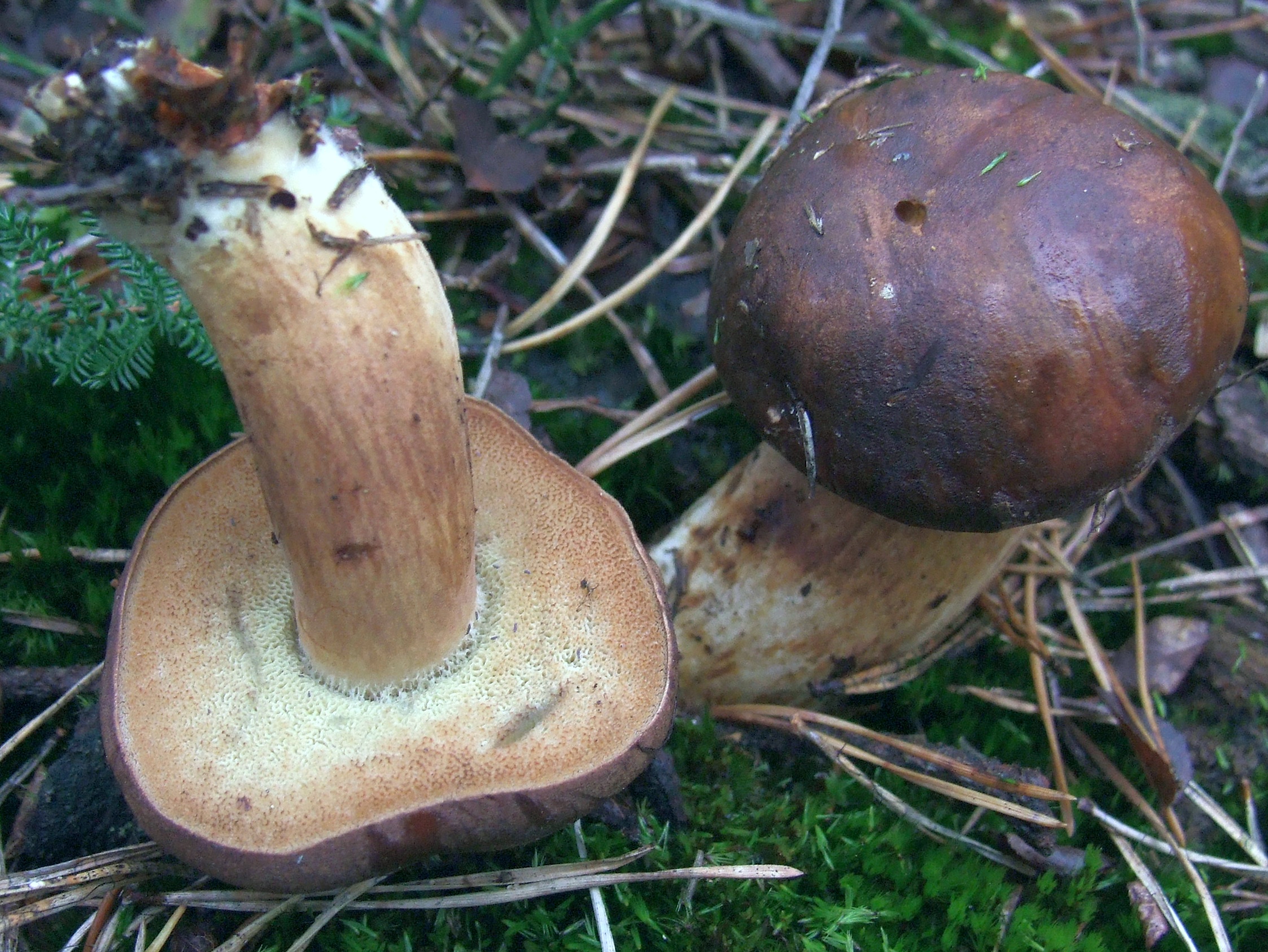
Jerzy Opioła · CC BY-SA 4.0
Korekta kolorów: Selso, 2020 (wersja źródłowa).
Photo source · CC BY-SA 4.0Imleria badia
Key differences
The matt bolete (Xerocomellus pruinatus) can resemble the bay bolete in cap coloration.
Xerocomellus chrysenteron
Key differences
The red cracking bolete (Xerocomellus chrysenteron) is characterized by a cap that cracks distinctly and deeply into patches, revealing reddish flesh beneath, as well as smooth spores. The matt bolete usually has a uniform, uncracked cuticle, and its spores are covered with fine longitudinal striations. Furthermore, the Czech atlas emphasizes that X. chrysenteron appears earlier in the season, already in early summer.
Xerocomellus porosporus
Key differences
Xerocomellus porosporus has characteristically truncate spore apices and, according to the Czech atlas, lacks such distinct red coloration on the stem. The mere similarity of the brown caps does not settle this comparison in the field.
Xerocomellus ripariellus
Key differences
Reddish forms of the matt bolete may resemble Xerocomellus ripariellus. Klofac points out that X. pruinatus is distinguished by yellow, rarely bluing flesh, a velvety and rarely cracking cap, slightly larger spores, and the structure of the pileipellis with variable terminal cells that are generally narrower than the subterminal cells lying beneath them.
FAQ
Is the matt bolete an edible mushroom?
Yes, the matt bolete is a valuable edible mushroom. It is prized for its dense, firm flesh, which is rarely infested with maggots, and its pleasant, mild taste. It is suitable for many dishes.
How can you identify the matt bolete?
It is characterized by a dark brown cap with a matte, frosted-like bloom in young specimens. The stem is yellowish in the upper part and reddish at the bottom, while the tubes are yellow, turning slightly blue.
Where and when does the matt bolete grow?
It grows in deciduous and mixed forests, particularly under beeches and oaks, less frequently under spruces. Fruit bodies appear from summer through late autumn, often even into November.
Does the flesh of the matt bolete change color when cut?
The flesh upon cutting is creamy or yellowish. In the cap and upper part of the stem, it may turn faintly blue upon exposure to air, while in the lower part of the stem it can be reddish.
Which mushrooms can the matt bolete be confused with?
It is most often confused with the red cracking bolete or the ribbed bolete. The red cracking bolete has a more strongly cracking cuticle with a reddish background, whereas the matt bolete has a dark and frosted cap.
Is the matt bolete suitable for drying?
Yes, it is excellent for drying. Thanks to the dense structure of its flesh, it retains good form, and after drying it has an intense, pleasant aroma, making it a great addition to sauces and soups.
Can the matt bolete be consumed raw?
No. According to sanitary-epidemiological recommendations, wild mushrooms should not be eaten raw. The matt bolete is an edible species, but for general sanitary reasons, it requires heat treatment.
What taste and smell does the matt bolete have?
This mushroom has a mild, pleasant taste and a delicate, mushroom aroma. It has no bitter or acidic aftertaste, making it a very versatile cooking ingredient.
Enthusiast
Names and history
- Obowiązująca nazwa krajowa
- suchogrzybek oprószony
- Obowiązująca nazwa taksonomiczna
- Xerocomellus pruinatus (Fr.) Šutara
- Inne nazwy krajowe
- podgrzybek aksamitny
- Dawne nazwy krajowe
- borowik oprószony, grzyb omszony, podgrzybek oprószony
Home observations
Spore print
- Required materials
- fresh, mature cap, sheet of paper, glass or small bowl, water
- Time
- 2–24 h
- Procedure
- Place the cap underside down on the paper.
- Add a drop of water on top and cover.
- After 2–24 hours, lift the cap and examine the deposit.
What to look for
- Spore print colour
- Spore print colour: green. Spore print colour: brown.
- Spore print shade
- Spore print shade: brownish-olive. Spore print shade: olive-brown.
Under the microscope
- Required materials
- light microscope, microscope slide and cover slip, needle, water, spores from print
- Procedure
- Transfer a small amount of the spore print onto the slide using a needle.
- Add a drop of water, cover with a cover slip, and examine the spores.
What to look for
- Spore shape
- Spore shape: ellipsoid. Spore shape: fusiform.
- Spore dimensions
- Length 10–15.7 µm, width 4–6.4 µm.
Compare dimensions after calibrating the microscope scale.
Ecology and distribution
- Soil calcareousness
- Soil calcareousness: calcareous.
- Broadleaf trees in the vicinity
- Broadleaf trees in the vicinity: oak or beech.
Broadleaf trees in the vicinity: beech or oak. - Coniferous trees in the vicinity
- Coniferous trees in the vicinity: spruce.
- Mycorrhizal hosts
- General species association: Mycorrhizal hosts: Fagus (general partner list).
General species association: Mycorrhizal hosts: Quercus (general partner list).
History and culture
The taxonomic history of the matt bolete reflects many years of research into the systematics of boletes and revisions of traditionally defined genera. The species was originally described in the 19th century by Elias Magnus Fries under the name Boletus pruinatus, and later in mycological literature, it was commonly placed in the genus Xerocomus as Xerocomus pruinatus.
A breakthrough moment in the taxonomic history of this taxon was the work of Czech mycologist Josef Šutara, published in 2008 in the journal "Czech Mycology" (vol. 60, issue 1, pp. 49–50). In this publication, Šutara segregated the new genus Xerocomellus based on a thorough analysis of micro- and macromorphological characteristics, transferring the species in question to this genus and establishing the new nomenclatural combination Xerocomellus pruinatus (Fr.) Šutara. This change has become an enduring reference point in contemporary bolete taxonomy in Europe.
Edibility and safety
The matt bolete is an edible mushroom characterized by firm and dense flesh. The primary condition for its safe consumption is subjecting the fruit bodies to thorough heat treatment before eating, such as by boiling or frying. Consuming raw or undercooked fruit bodies may lead to gastrointestinal discomfort. Only young, healthy, and fresh specimens should be used for culinary purposes.
A significant risk factor is the rapid aging of fruit bodies and the susceptibility of their tissues to spoilage. Old, overripe, waterlogged specimens or those showing any signs of mold are unfit for consumption. Additionally, as with other boletes, the fruit bodies may accumulate contaminants from the soil and surrounding environment; for this reason, gathering specimens growing in the immediate vicinity of heavily trafficked roads and in heavily industrialized areas is not recommended.
In terms of flavor qualities, the flesh of the matt bolete is characterized by a mild taste free of bitterness and a delicate, typically mushroom aroma, which makes it suitable for use in the kitchen as a domestic edible mushroom.
Habitat and occurrence
The matt bolete (Xerocomellus pruinatus) occurs in Europe, primarily in deciduous and mixed forests, and less frequently in pure coniferous stands. Sites of this species are also recorded in parks and landscaped plantings. The fungus forms mycorrhizal associations with broadleaved trees, especially European beech, oaks, and European hornbeam, as well as with conifers, including Norway spruce. It prefers fresh, nutrient-rich, loamy, and sandy-loamy soils, where fruit bodies grow singly or in groups on forest litter, among fallen leaves, and moss cushions.
The geographic range of the species covers Europe, including Central and Western Europe, extending north to the southern regions of the Scandinavian Peninsula and south toward the Mediterranean basin.
Under Poland's climatic conditions, the fruiting season falls between September and November (months IX–XI), corresponding to the documented emergence of fruit bodies in Central European phenology. The occurrence of the species in a given region depends primarily on the presence of favorable deciduous and mixed forest stands and moist loamy soils.
Researcher
Edibility and culinary use
Ustalenia źródłowe: jadalność, spożycie i wykorzystanie
- Edibility
- Edible — źródło oznaczenia
Česká mykologická společnost, Xerocomellus pruinatus, hřib sametový · Kuchyňské využití: Hřib sametový je dobrá jedlá houba.
- Culinary rating
- Tasty
Česká mykologická společnost, Xerocomellus pruinatus, hřib sametový · Kuchyňské využití: dobrá jedlá houba, z druhů rodu Xerocomellus patrně nejchutnější. Source opinion; not a formal quality class or larval-infestation claim.
Scientific names and taxonomy
- Current scientific name
Xerocomellus pruinatus (Fr.) Šutara
- Former scientific names
- 1835 — Boletus pruinatus Fr.
1886 — Versipellis pruinata (Fr.) Quél.
1888 — Xerocomus pruinatus (Fr.) Quél.
1898 — Suillus pruinatus (Fr.) Kuntze
1992 — Boletellus pruinatus (Fr.) Klofac & Krisai[17]
Suchogrzybek oprószony należy do rodziny borowikowatych (Boletaceae) w rzędzie borowikowców (Boletales). Nazwa naukowa taksonu to Xerocomellus pruinatus (Fr.) Šutara. Kombinację tę opublikował w 2008 r. Josef Šutara pod numerem rejestracyjnym MB511898, przenosząc do nowego rodzaju gatunek opisany pierwotnie w 1835 r. przez Eliasa Friesa jako Boletus pruinatus Fr. Takie samo autorstwo bazonimu podaje baza Species Fungorum. W części opracowań pojawia się zapis „Fr. & Hök”, jednak Oolbekkink uzasadniał przypisanie pierwotnego opisu samemu Friesowi.
[17] [19] [13] [7] [15] [18]Wcześniejsze kombinacje oparte na tym samym epitecie gatunkowym obejmują Versipellis pruinata (Fr.) Quél. z 1886 r., Xerocomus pruinatus (Fr.) Quél. z 1888 r., Suillus pruinatus (Fr.) Kuntze z 1898 r. oraz Boletellus pruinatus (Fr.) Klofac & Krisai z 1992 r. Szereg ten tłumaczy, dlaczego w dawniejszej literaturze mikologicznej ten sam grzyb bywał umieszczany w różnych rodzajach.
[17] [19] [13] [7] [15] [18]W rejestrze Species Fungorum do synonimiki gatunku włączono także nazwy Boletus subtomentosus var. pascuus Pers. (1825), Boletus pascuus (Pers.) Corda (1841), Xerocomus pascuus (Pers.) Bon (1985 [1984]), Boletellus pruinatus var. pascuus (Pers.) Klofac & Krisai (1992) oraz Boletellus pruinatus f. luteocarnosus Klofac & Krisai (1992). Stanowią one historyczne przyporządkowania taksonomiczne w rangach odmiany i formy.
[17] [19] [13] [7] [15] [18]Taxon scope and identification limitations
Identyfikacja mikroskopowa gatunków z rodzaju Xerocomellus opiera się przede wszystkim na strukturze skórki kapelusza oraz mikrorzeźbie zarodników. Zarodniki suchogrzybka oprószonego pokryte są drobnymi, podłużnymi prążkami, jednak autorzy Fungi of Temperate Europe zaznaczają, że struktury te pozostają niewidoczne w standardowej mikroskopii świetlnej. Wobec tego ani sama ciemna barwa kapelusza, ani intensywne sinienie nie wystarczają do jednoznacznego oznaczenia bez weryfikacji pozostałych cech.
[7] [6] [13] [9] [14] [8]Šutara badał głównie skrawki wysuszonych owocników (cięte ręcznie żyletką), z użyciem 3–10% NH4OH, niekiedy z dodatkiem czerwieni Kongo oraz odczynnika Melzera lub w czystym NH4OH; świeży materiał analizował w wodzie, odczynniku Melzera lub wodnym roztworze czerwieni Kongo, stosując również mikroskop SEM. Diagnoza całego rodzaju Xerocomellus obejmuje grubość skórki (80–)120–350(–500) µm, rurki do 10(–14) mm i kanciaste pory o średnicy 1–2,5 mm. Pileipellis tworzą równoległe łańcuszki komórek, których końce leżą na zbliżonym poziomie; u starszych owocników układ ten może się rozluźniać. Są to cechy i wymiary diagnozy rodzajowej, odrębne od indywidualnych pomiarów gatunkowych X. pruinatus.
[19]Cechy mikroskopowe rodzaju Xerocomellus obejmują ponadto słabo żelatynizującą tramę hymenoforu typu pośredniego z odległościami między strzępkami 2–4(–8) µm, przeważnie wrzecionowato-butelkowate pleurocystydy o ściankach do 0,6 µm, brak lub zredukowaną do 30(–40) µm boczną warstwę trzonu, płodną warstwę kaulohymenialną na trzonie oraz brak sprzążek na strzępkach. Brak osłony częściowej i pierścienia stanowi stałą cechę całego rodzaju.
[19] [6]W rewizji taksonomicznej Šutary uwzględniono 15 kolekcji zielnikowych tego gatunku: JŠ 078, 302, 336, 337, 341, 345, 1836, 1963, 1965, 1986, 2112, 4053, 4397, 4398 oraz 4399.
[19]Abstrakt pracy Peintner, Ladurner i Simoniniego z 2003 r. opisuje analizę 24 sekwencji rDNA-LSU wykazującą odrębne linie ośmiu taksonów, w tym pruinatus. Pomocne w diagnozie były cechy zarodników, komórki końcowe skórki oraz charakterystyczne strzępki typu pruinatus (pruinatus-hyphae). Autorzy wykazali jednak wielokrotne powstawanie w toku ewolucji podobnego prążkowania zarodników, ściętych wierzchołków oraz strzępek tego typu. Liczba 24 sekwencji odnosi się do całego badania obejmującego osiem taksonów.
[14]Badania Franka i współpracowników z 2020 r. nad północnoamerykańskimi przedstawicielami rodzaju Xerocomellus uwzględniały sekwencje referencyjne europejskiego X. pruinatus z bazy GenBank (LSU: AF514825 z okazu IB19961055, AF050644 oraz AF402140), wskazując na konieczność ostrożnej weryfikacji oznaczeń w publicznych repozytoriach sekwencji.
[8]Laboratory diagnostics
Dane ustrukturyzowane
Pileipellis
- Incrustation of cuticle hyphae
- Incrustation of cuticle hyphae: present. [6]
Incrustation of cuticle hyphae: present. [13] - Structure of the pileipellis
- Structure of the pileipellis: palisadoderm. [6]
Structure of the pileipellis: trichoderm. [13] - Pileipellis terminal cell width
- Pileipellis terminal cell width: 5.5–15.5 µm. [13]
Reactions with reagents
- Reaction to alkaline reagents
- Reaction to alkaline reagents: no colour change.
Reaction to alkaline reagents: no colour change.
Spores
- Spore length
- Spore length: 11–15 µm. [6]
Spore length: 10–14.5 µm. [9]
Spore length: 11–14 µm. [3]
Spore length: 11.5–14 µm. [7]
Spore length: 11.5–15 µm. [18]
Spore length: 11.4–14.9 µm. [18]
Spore length: 12.3–15.3 µm. [18]
Spore length: 12–15.7 µm. [18] - Form of spore ornamentation
- Form of spore ornamentation: striate. [7]
Form of spore ornamentation: striate. [6]
Form of spore ornamentation: striate. [9]
Form of spore ornamentation: striate. [13] - Direction of spore ornamentation
- Direction of spore ornamentation: longitudinal. [7]
Material and research methods
- Results as recorded in the sources — Spore length
- 11,5–15 µm [18]
10–14,5 µm [9]
12–15,1 µm (skrajnie do 15,7 µm) [18]
11–15 µm [6]
11,5–14 µm [7]
11,4–14,9 µm [18]
12,3–15,3 µm [18]
11–14 µm [3] - Results as recorded in the sources — Spore width
- 4,5–5,5 µm [9] [7]
4,2–5,7 µm [18]
4,4–5,4 µm (skrajnie do 6 µm) [18]
4,5–6 µm [6]
4,9–6,4 µm [18]
4,3–5,8 µm [18]
4–5 µm [3]
Metody i zapisy źródłowe — Source cap-cuticle microscopy; source-specific terminology and scantiness are not merged into one universal organisation.
Inkrustacja strzępek skórki; Organizacja skórki kapelusza — zapis źródłowy: „Species Description: Pileipellis palisadoderm of septate hyphae of cylindrical, incrusted cells.” [6]
Metody i zapisy źródłowe — Source cap-cuticle microscopy; source-specific terminology and scantiness are not merged into one universal organisation.
Inkrustacja strzępek skórki; Organizacja skórki kapelusza — zapis źródłowy: „Printed p.265 section9: somewhat irregular trichoderm; terminal cells(5.5–)8–14(–15.5)µm broad; incrustations scarce.” [13]
Szerokość komórki końcowej skórki kapelusza — zapis źródłowy: „Terminal cells(5.5–)8–14(–15.5)µm broad.” [13]
Metody i zapisy źródłowe — ČMS species description; material, voucher, measurement count and medium unreported.
Długość zarodnika — zapis źródłowy: „11.5–14 × 4.5–5.5 µm; first axis.” [7]
Forma ornamentacji zarodnika — zapis źródłowy: „jemně podélně rýhované” [7]
Kierunek ornamentacji zarodnika — zapis źródłowy: „podélně rýhované” [7]
Kształt zarodnika — zapis źródłowy: „eliptické nebo vřetenovité” [7]
Szerokość zarodnika — zapis źródłowy: „11.5–14 × 4.5–5.5 µm; second axis.” [7]
Metody i zapisy źródłowe — Published species description; unreported individual material and numerical protocol.
Długość zarodnika — zapis źródłowy: „Species Description: Spores11–15×4.5–6µm, striate. First axis.” [6]
Forma ornamentacji zarodnika — zapis źródłowy: „Species Description: Spores11–15×4.5–6µm, striate.” [6]
Szerokość zarodnika — zapis źródłowy: „Species Description: Spores11–15×4.5–6µm, striate. Second axis.” [6]
Metody i zapisy źródłowe — Published species description; unreported individual material and numerical protocol.
Długość zarodnika — zapis źródłowy: „Printed p.579 couplet73: spores10–14.5×4.5–5.5µm, very finely striated, not visible in a standard light microscope. First axis.” [9]
Forma ornamentacji zarodnika — zapis źródłowy: „Printed p.579 couplet73: spores10–14.5×4.5–5.5µm, very finely striated, not visible in a standard light microscope.” [9]
Szerokość zarodnika — zapis źródłowy: „Printed p.579 couplet73: spores10–14.5×4.5–5.5µm, very finely striated, not visible in a standard light microscope. Second axis.” [9]
Metody i zapisy źródłowe — Focal Xerocomellus pruinatus row, PDFpage81; individual microscopic material unreported.
Długość zarodnika; Szerokość zarodnika — zapis źródłowy: „11–14×4–5µm, paired source axes.” [3]
Metody i zapisy źródłowe — 19Nov2021, PuszczaBukowa Węglino, leg./det.GrażynaDomian; mean13.5×5.5µm,Qmean2.4.
Długość zarodnika; Szerokość zarodnika — zapis źródłowy: „19Nov2021, PuszczaBukowa Węglino, leg./det.GrażynaDomian; mean13.5×5.5µm,Qmean2.4. See original paired dimensions.” [18]
Iloraz długości i szerokości zarodnika — zapis źródłowy: „19Nov2021, PuszczaBukowa Węglino, leg./det.GrażynaDomian; mean13.5×5.5µm,Qmean2.4. Source Q range, not recomputed from unpaired dimension endpoints.” [18]
Metody i zapisy źródłowe — 25Oct2023, BukoweZdroje, leg./det.GrażynaDomian; mean12.9×4.8µm,Qmean2.7.
Długość zarodnika; Szerokość zarodnika — zapis źródłowy: „25Oct2023, BukoweZdroje, leg./det.GrażynaDomian; mean12.9×4.8µm,Qmean2.7. See original paired dimensions.” [18]
Iloraz długości i szerokości zarodnika — zapis źródłowy: „25Oct2023, BukoweZdroje, leg./det.GrażynaDomian; mean12.9×4.8µm,Qmean2.7. Source Q range, not recomputed from unpaired dimension endpoints.” [18]
Metody i zapisy źródłowe — 30Oct2023, BuczynoweWąwozy, leg./det.GrażynaDomian; mean13.1×4.9µm,Qmean2.7.
Długość zarodnika; Szerokość zarodnika — zapis źródłowy: „30Oct2023, BuczynoweWąwozy, leg./det.GrażynaDomian; mean13.1×4.9µm,Qmean2.7. See original paired dimensions.” [18]
Iloraz długości i szerokości zarodnika — zapis źródłowy: „30Oct2023, BuczynoweWąwozy, leg./det.GrażynaDomian; mean13.1×4.9µm,Qmean2.7. Source Q range, not recomputed from unpaired dimension endpoints.” [18]
Metody i zapisy źródłowe — 4Nov2023, TrawiastaBuczyna, leg./det.GrażynaDomian; mean13.5×5.0µm,Qmean2.7.
Długość zarodnika; Szerokość zarodnika — zapis źródłowy: „4Nov2023, TrawiastaBuczyna, leg./det.GrażynaDomian; mean13.5×5.0µm,Qmean2.7. See original paired dimensions.” [18]
Iloraz długości i szerokości zarodnika — zapis źródłowy: „4Nov2023, TrawiastaBuczyna, leg./det.GrażynaDomian; mean13.5×5.0µm,Qmean2.7. Source Q range, not recomputed from unpaired dimension endpoints.” [18]
Metody i zapisy źródłowe — Source B.pruinatus description, with collections Bos8335 (NL) and Oolbekkink207 (DE) listed, without assigning one individual SEM image to either voucher.
Metoda źródłowa. Technika: skaningowa mikroskopia elektronowa (SEM). Opis metody w źródle: Source methods p.246: gold-coated stubs, 15kV JEOL JSM-35; source spore prints or tube fragments, not an inferred focal specimen preparation.
Forma ornamentacji zarodnika — zapis źródłowy: „Faint to distinct striae with SEM.” [13]
Occurrence verification — scope and limitations
Materiał: Zbiór z Puszczy Bukowej w województwie zachodniopomorskim; status zweryfikowany potwierdzono w osobnym publicznym wpisie GRej.
Zakres odczytu: Fragmenty źródeł.
Odczytano fragment źródła; pełny tekst i metody nie zostały zweryfikowane.
Data przeglądu: 21.10.2026.
Ograniczenia: Źródłowe oznaczenie i zakres pojęcia zachowano. Nie zadeklarowano nowego badania okazów ani odczytu wszystkich publikacji cytowanych przez źródło.
[18]Scope of the review
Before completion, the record requires completion or settlement.
- Search for occurrence regions requires reconciliation.
- Connections of earlier findings require re-examination.
- New sources require accounting for information encountered during the review.
Key substances and their effects
W badaniu korzeni jodły pobrano w październiku 2012 r. po 16 kostek gleby 15 × 15 × 15 cm z czterech stanowisk: Kartuzy, Lipusz, Osusznica i Sławno (łącznie 64 monolity). Analizowano drobne korzenie poniżej 2 mm w trzech losowych podpróbach po około 250 wierzchołków. Oznaczenie ITS suchogrzybka oprószonego KP230473 wykazało 99% zgodności z UDB000481 przy kryterium badania przekraczającym 98% na odcinku co najmniej 450 pz. Udział gatunku wynosił odpowiednio 1,5 ± 0,3%, 1,7 ± 0,3%, 0,9 ± 0,2% oraz 2,0 ± 0,1% (średnia ± błąd standardowy, SE).
[16]W badanych drzewostanach jodłowych odczyn gleby mierzony w wodzie wynosił w poziomie organicznym (O) odpowiednio 3,9; 3,9; 3,9 i 4,0, a w poziomie mineralnym (A): 4,0; 4,6; 4,6 oraz 4,3. Typy gleb sklasyfikowano jako glebę kwaśną brunatną (Kartuzy, Lipusz, Sławno) oraz glebę płową (Osusznica).
[16]W doświadczeniu Zavišić i współautorów materiał buka z zasobniejszego w fosfor Bad Brückenau i ubogiego Unterlüß przeniesiono w październiku 2014 r. do wspólnego ogrodu w Getyndze, gdzie rósł do lipca 2015 r. Badano 40 roślin, po 10 na wariant i pochodzenie gleby; 20 otrzymało znacznik 33P, a 20 stanowiło kontrolę bez znacznika. Po miesiącu aklimatyzacji, 20 listopada 2014 r., zastosowano jednorazową dawkę 795 mg P na roślinę. Mikoryzy identyfikowano na podstawie sekwencji ITS przy kryterium zgodności ponad 97% i wyniku dopasowania (score) ponad 900 bitów, co pozwoliło rozpoznać 14 z 16 morfotypów.
[21]Po nawożeniu fosforem udział mikoryz suchogrzybka oprószonego zmniejszył się w materiale z gleby zasobnej w fosfor. Wynik testu ANOSIM p = 0,385 oznaczał jednak brak statystycznie istotnej zmiany struktury całej zbiorowości grzybów mikoryzowych w tej glebie, a nie oddzielny test liczebności samego gatunku. Nie rozpoznano mechanizmu pobierania fosforu przez izolowany szczep, a nierozpoznany morfotyp MT31 stanowił osobną pozycję. Wyjściowe pH gleby wynosiło 3,8 w Bad Brückenau i 3,5 w Unterlüß, co opisuje warunki miejsc pobrania, a nie optimum gatunku.
[21]W badaniu Kautmanovej i współautorów materiał zbierano na słowackich terenach pogórniczych w latach 2022–2024, a oznaczenia oparto na morfologii i sekwencjach ITS. Oczyszczone owocniki krojono i suszono do stałej masy w temperaturze do 60 °C; małe mielono w całości, a z dużych pobierano równe części kapelusza, trzonu i hymenoforu, uzyskując co najmniej 2 g homogenizowanego proszku. Do analizy ICP-MS mineralizowano 1 g suchego materiału kwasem azotowym, a następnie wodą królewską. Trzy wykonane powtórzenia miały charakter analityczny, a liczba niezależnych owocników gatunku nie została w pracy wyszczególniona.
[11]W materiale ze Słowacji stężenie kadmu w owocnikach z Gelnicy wynosiło 7,82 mg/kg suchej masy (wskaźnik THQ 0,8), a ze Zlatej Idki 3,95 mg/kg s.m. (THQ 0,4). Zawartość ołowiu wynosiła odpowiednio 4,86 mg/kg s.m. (THQ 0,1) w Gelnicy oraz 1,77 mg/kg s.m. przy współczynniku bioakumulacji BCF 0,001 w Zlatej Idce.
[11]W Gelnicy odnotowano rtęć na poziomie 14,23 mg/kg suchej masy, z modelowym wskaźnikiem zagrożenia THQ 7,6 oraz współczynnikiem biokoncentracji BCF 2,14, a także mangan w stężeniu 974 mg/kg suchej masy. THQ stanowi wskaźnik modelowy, a nie liczbę klinicznych zatruć, natomiast wysoki poziom manganu nie oznaczał jego bioakumulacji z podłoża (dla manganu publikacja wskazuje brak akumulacji z gleby). Wszystkie wartości dotyczą zbadanego suchego materiału bez przeliczania na świeżą masę i bez uogólniania na owocniki z innych obszarów.
[11]Habitat and occurrence
Habitat, region and season
Gatunek wyrasta na ziemi w lasach liściastych, mieszanych i iglastych oraz w parkach. Assyov wymienia jako głównych partnerów mikoryzowych buki, a rzadziej dęby, natomiast autorzy czescy podają buki, dęby i świerki. W polskich rejestrach stanowiska obejmują kwaśne i żyzne buczyny, leśną ściółkę, obrzeża drzewostanów, pobliża nadrzecznych łęgów, a także ciepłe, piaszczyste murawy na skraju lasu dębowego. Sama obecność sosny, brzozy czy grabu w opisie drzewostanu nie stanowi bezpośredniego dowodu na tworzenie mikoryzy z tymi gatunkami drzew.
[7] [6] [18] [20] [9]Analizy molekularne korzeni jodły pospolitej (Abies alba) na Pomorzu potwierdziły obecność tego grzyba w ektomikoryzach na czterech stanowiskach zlokalizowanych poza naturalnym zasięgiem jodły. W badaniach w Niemczech zidentyfikowano go również w mikoryzach buka zwyczajnego (Fagus sylvatica). Wyniki te uzyskano bezpośrednio z sekwencjonowania ITS korzeni, co stanowi niezależne potwierdzenie laboratoryjne relacji mikoryzowej.
[16] [21]Assyov uznaje gatunek za szeroko rozpowszechniony w Europie, zaznaczając problem mylenia go z pokrewnymi taksonami. Czeski atlas określa go jako obfity, a europejski klucz jako pospolity. Władysław Wojewoda w 2003 r. przytaczał dawne stwierdzenie z Puszczy Białowieskiej oraz kategorię PL–Ex, a atlas leśny z 2021 r. odnotowywał historyczną ocenę uznającą go za gatunek bardzo rzadki, znany niemal wyłącznie z tego rejonu. Liczne współczesne udokumentowane stanowiska w Polsce (w tym późniejsze zgłoszenia pomorskie i z innych rejonów) wskazują jednak na znacznie szersze występowanie.
[20] [18] [6] [7] [9]W Wielkiej Brytanii zestawienie z 2013 r. wykazało obecność gatunku w 10 kwadratach siatki 1 × 1 km w latach 1913–1962 oraz w ponad 200 kwadratach w latach 1963–2012, kwalifikując go do kategorii najmniejszej troski (LC). Szacunek ponad 2000 dojrzałych osobników stanowi wynik modelowania opartego na bazach obserwacji, a nie bezwzględną liczbę zliczonych owocników.
[10]Potwierdzony zasięg występowania
Wszystkie obszary potwierdzone w zapisanych danych źródłowych.
| Kraj | Regiony i stanowiska | Owocnikowanie | Sources |
|---|---|---|---|
| Poland | West Pomeranian Voivodeship | IX–XI |
|
Sources
- grenoble 2015
- Boris Assyov, Xerocomus pruinatus
- Česká mykologická společnost, Xerocomellus pruinatus, hřib sametový
- src-frank 2020
- Læssøe T., Petersen J.H., Frøslev T.G., Heilmann-Clausen J. 2025. Keys to the fungi of Temperate Europe,8XI 2025
- src-jncc 2013
- src-kautmanova 2026
- Oolbekkink G.T. 1991. The taxonomic value of the ornamentation of spores in The Xerocomus-group of Boletus. Persoonia14(3): 245–273
- src-peintner 2003-abstract
- src-ptmyk 2021
- src-rudawska 2016
- Species Fungorum, rekord 511898
- Marek Snowarski, Tx236, Xerocomellus pruinatus
- src-sutara 2008
- Wojewoda W. 2003. Checklist of Polish larger Basidiomycetes. Biodiversity of Poland7
- src-zavisic 2018