
Inedible

Set your location to see data for your region.


The inkstain bolete is a mushroom from the family Boletaceae, found in deciduous, coniferous, and mixed forests, as well as in parks. It produces a cap coloured brown to olive-brown and yellow pores. The stipe may be cylindrical or tapering towards the base, yellow at the top and reddish-brown in the lower part. A characteristic feature is the exceptionally rapid and intense bluing of the pores and flesh when bruised. Formerly considered an edible mushroom, it is currently advised against because studies have demonstrated arsenic accumulation.
Identification, habitat, and key differences.
Forager
Fruitbody structure
- Fruit body form
- Fruit body form: Cap, stipe.
- Consistency of the entire fruitbody
- Consistency of the entire fruitbody: fleshy.
Cap
- Cap shape
- in young fruit bodies: Cap shape: hemispherical.
in mature fruit bodies: Cap shape: convex.
in old fruit bodies: Cap shape: flat.
in mature fruit bodies: Cap shape: flat. - Cap surface relief
- in mature fruit bodies: Cap surface relief: smooth.
- Cap covering
- in young fruit bodies: Cap covering: suede-like.
Cap covering: silky.
in young fruit bodies: Cap covering: powdery.
in young fruit bodies: Cap covering: felty.
in mature fruit bodies: Cap covering: glabrous. - Cap surface sheen
- in young fruit bodies: Cap surface sheen: matt.
- Cap stickiness and sliminess
- when wet: Cap stickiness and sliminess: sticky.
Cap stickiness and sliminess: sticky. - Surface cracking pattern
- Surface cracking pattern: cracked, pattern unspecified.
- Cap colour
- Cap colour: brown.
Cap colour: crimson.
Cap colour: dark brown.
Cap colour: gray-green.
Cap colour: olive.
Cap colour: olive-brown.
Cap colour: pink.
Cap colour: reddish brown.
Cap colour: yellow.
Cap colour: yellow-brown.
Cap colour: yellowish.
Cap colour: dirty dark brown. - Margin orientation
- Margin orientation: straight.
in old fruit bodies: Margin orientation: turned upwards.
in young fruit bodies: Margin orientation: curved inward. - Cap margin outline
- Cap margin outline: lobed, wavy.
- Cap margin profile
- Cap margin profile: acute.
- Cap colour reaction
- Cap colour reaction: black.
- Cap diameter
- The cap measures 40–120 mm in diameter.
Underside of the cap
- Hymenophore type
- Hymenophore type: tubes.
Tubes at the stem
- Hymenophore attachment to the stipe
- Hymenophore attachment to the stipe: attached to the stipe (tubes).
- Decurrence of the hymenophore on the stem
- in mature fruit bodies: Decurrence of the hymenophore on the stem: decurrent (tubes).
- Pore shape
- Pore shape: angular.
Pore shape: irregular. - Pore colour after damage
- Pore colour after damage: blue-black.
- Tube color after damage
- Tube color after damage: blue-black.
- Natural pore colour
- in mature fruit bodies: Natural pore colour: yellow-olive.
Natural pore colour: yellow.
Natural pore colour: orange. - Natural tube colour
- Natural tube colour: lemon yellow.
in mature fruit bodies: Natural tube colour: yellow-olive.
Stem and base
- Stipe shape
- Stipe shape: cylindrical or tapering towards the base.
Stipe shape: fusiform. - Stipe base
- Stipe base: cylindrical.
Stipe base: tapering. - Stipe colour
- in the upper part of the stipe: Stipe colour: vivid yellow.
in the lower part of the stipe: Stipe colour: reddish brown. - Stipe surface relief
- in the upper part of the stipe: Stipe surface relief: reticulate.
- Stipe surface covering
- in the lower part of the stipe: Stipe surface covering: powdery.
- Color of stem surface upon damage
- Color of stem surface upon damage: blue-black (stipe surface).
- Stipe length
- Stipe length: 40–100 mm.
- Stipe diameter
- Stipe diameter: 5–25 mm.
- Stipe curvature
- Stipe curvature: curved.
Flesh
- Flesh colour
- Flesh colour: yellow.
in old fruit bodies: Flesh colour: red (stipe flesh).
Flesh colour: whitish or yellowish. - Flesh firmness
- in young fruit bodies: Flesh firmness: firm.
- Flesh taste
- Flesh taste: sour.
Flesh taste: weak.
Flesh taste: sweet. - Flesh colour after damage
- Flesh colour after damage: blue-black (cap flesh).
Flesh colour after damage: blue-black (stipe flesh). - Flesh odour
- Flesh odour: weak.
Where and when it grows
- Habitat
- Habitat: deciduous woodland or coniferous woodland.
Habitat: deciduous woodland, mixed woodland, park or garden.
Habitat: deciduous woodland. - Substrate
- Substrate: soil.
- Fruit body growth pattern
- Fruit body growth pattern: solitary or in groups.
- Occurrence frequency
- Fruiting season
- Fruit bodies appear from June to November.
- Distribution area
- Distribution area: confirmed.
Edibility and culinary use
- Edibility
- Inedible
Law
- Protection
- In Poland, the inkstain bolete is not subject to legal species protection.
- Commercial trade
- The inkstain bolete does not appear on the Polish list of mushrooms permitted for trade and the production of processed goods.
What it can be confused with
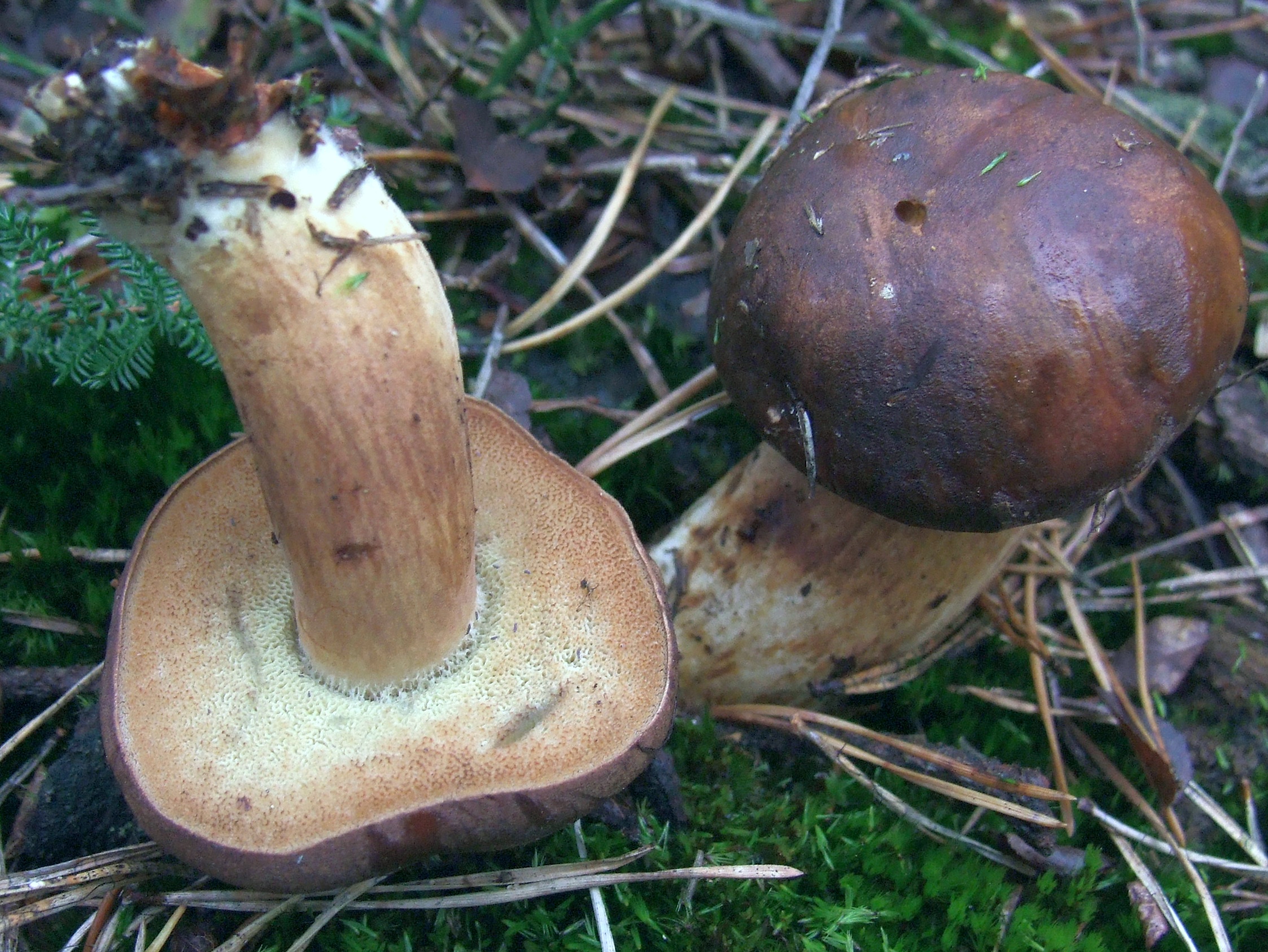
Jerzy Opioła · CC BY-SA 4.0
Korekta kolorów: Selso, 2020 (wersja źródłowa).
Photo source · CC BY-SA 4.0Imleria badia
Key differences
The inkstain bolete (Cyanoboletus pulverulentus) reacts to touch and damage with an immediate, rapid, and intense discolouration of all parts of the fruit body to a deep navy-black colour. In Imleria badia, the bluing is less uniform and is concentrated primarily above the tubes and in the upper part of the stipe.
FAQ
Is the inkstain bolete an edible mushroom?
In old field guides it was considered edible, but today its consumption is advised against. Studies have demonstrated this species' ability to accumulate arsenic, which is why the authors of publications advise against its consumption.
Why is the consumption of this species advised against?
The reason is research findings demonstrating high accumulation of arsenic in the fruit bodies. For this reason, mycologists and authors of newer studies recommend refraining from harvesting it for culinary purposes.
What are the main identifying features of this mushroom?
Upon bruising or cutting, the pores and flesh turn dark blue very quickly. The tubes are yellow, and the stipe may be cylindrical or tapered at the base, yellow at the top and reddish-brown at the bottom.
In what habitats does this species occur?
It grows in deciduous, coniferous, and mixed forests. It can also be encountered outside the forest, for example in parks and gardens.
In which months can it be found?
In the literature, the reported occurrence period of fruit bodies falls between June and November. Different sources indicate narrower ranges, depending on local weather conditions.
How does it differ from the bay bolete?
The inkstain bolete turns blue much faster and more intensely, adopting a dark shade. It also has yellow pores and a stipe that is yellow at the apex and reddish-brown towards the bottom, with possible fine structure at the top.
What is the former Polish name of this species?
Formerly in Polish literature, this species functioned under the name borowik klinowotrzonowy. The current name sinoborowik klinowotrzonowy is recommended by the Polish Mycological Society (PTMyk).
Enthusiast
Underside of the cap
- Tube length
- Tube length: up to 15 mm.
Names and history
- Obowiązująca nazwa krajowa
- sinoborowik klinowotrzonowy
- Obowiązująca nazwa taksonomiczna
- Cyanoboletus pulverulentus (Opat.) Gelardi, Vizzini & Simonini (2014)
- Inne nazwy krajowe
- Inkstain Bolete
- Dawne nazwy krajowe
- borowik klinowotrzonowy, borowik wrażliwy, borowik siniejący, borowik czerniejący, borowik omglony, podgrzybek omglony
Home observations
Spore print
- Required materials
- fresh, mature cap, sheet of paper, glass or small bowl, water
- Time
- 2–24 h
- Procedure
- Place the cap underside down on the paper.
- Add a drop of water on top and cover.
- After 2–24 hours, lift the cap and inspect the deposit.
What to look for
- Spore print colour
- Spore print colour: brown.
- Spore print shade
- Spore print shade: olive-brown.
Under the microscope
- Required materials
- light microscope, microscope slide and cover slip, needle, water, spores from print
- Procedure
- Transfer a small amount of the spore print onto the slide using a needle.
- Add a drop of water, cover with a cover slip, and examine the spores.
What to look for
- Spore shape
- Spore shape: nearly spindle-shaped. Spore shape: broadly ellipsoid.
- Spore surface
- Spore surface: smooth.
- Spore dimensions
- Length 10–15.5 µm, width 4–6 µm.
Compare dimensions after calibrating the microscope scale.
Ecology and distribution
- Soil reaction
- Soil reaction: acidic.
Soil reaction: neutral. - Broadleaf trees in the vicinity
- Broadleaf trees in the vicinity: Castanea sativa, Carpinus betulus, Fagus sylvatica, Quercus cerris, Quercus ilex, Quercus pubescens, Quercus robur or Tilia cordata.
Broadleaf trees in the vicinity: beech, lime tree, sweet chestnut or oak.
Broadleaf trees in the vicinity: Quercus coccifera, Quercus pyrenaica, Quercus suber, alder, birch or Ostrya. - Coniferous trees in the vicinity
- Coniferous trees in the vicinity: Pinus nigra, Pinus pinea, Pinus sylvestris, Cedrus libani or hemlock.
Coniferous trees in the vicinity: fir or Picea abies. - Shrubs in the vicinity
- Shrubs in the vicinity: Corylus avellana.
- Mycorrhizal hosts
- General species association: Mycorrhizal hosts: Castanea (general partner list).
General species association: Mycorrhizal hosts: Carpinus (general partner list).
General species association: Mycorrhizal hosts: Corylus (general partner list).
General species association: Mycorrhizal hosts: Fagus (general partner list).
General species association: Mycorrhizal hosts: Quercus (general partner list).
General species association: Mycorrhizal hosts: Tilia (general partner list).
General species association: Mycorrhizal hosts: Pinus (general partner list).
General species association: Mycorrhizal hosts: Cedrus (general partner list).
General species association: Mycorrhizal hosts: Tsuga (general partner list). - Mode of nutrition
- Mode of nutrition: ectomycorrhizal.
History and culture
The taxonomic history of the inkstain bolete is closely intertwined with the beginnings of modern mycology and carries a significant Polish historical accent. This species was first described scientifically in 1836 under the name Boletus pulverulentus by physician and mycologist Wilhelm Opatowski. The description appeared in his acclaimed dissertation devoted to boletes (Commentatio historico-naturalis de familia Fungorum Boletoideorum), published in Berlin. The specific epithet pulverulentus (Latin for powdery, frosted) referred to the delicate, as if powdered surface of young fruit bodies, which was also reflected in the Polish vernacular name.
For nearly two centuries, the taxon functioned in literature within the broadly defined genus Boletus. A breakthrough in its classification occurred in 2014, when Italian researchers Matteo Gelardi, Alfredo Vizzini, and Giampaolo Simonini carried out a phylogenetic revision of the boletes and erected the new genus Cyanoboletus, to which they transferred Opatowski's species. The name of the new genus was inspired by the spectacular, dark-blue color reaction of the oxidizing flesh. These changes reflect the evolution of systematic knowledge in European mycological literature.
Edibility and safety
The inkstain bolete (Cyanoboletus pulverulentus) is a conditionally edible mushroom that requires great caution from the consumer. In the raw state, it is toxic and causes severe irritation of the digestive tract, manifesting as abdominal pain, nausea, and vomiting. A specific health hazard associated with this species is its unique ability to hyperaccumulate arsenic from the substrate, including dimethylarsinic acid compounds.
A prerequisite for consumption is thorough heat treatment – boiling for at least 15–20 minutes and discarding the cooking water, or thorough frying. Despite preparation, consuming it in large portions or regularly is not recommended; it should be treated exclusively as a mushroom eaten occasionally.
The flesh of the raw mushroom has a pleasant, mild, slightly nutty flavor and a delicate aroma. Under pressure and when cut, it immediately bruises intensely blue, and then darkens, which after cooking yields an almost black color of the dish.
The approach to consumption varies regionally. In Poland, the Czech Republic, and Slovakia, it is often eagerly collected by experienced mushroom pickers as a tasty domestic mushroom, whereas in Western Europe (among others in Germany), due to concerns over arsenic content, health authorities advise caution and discourage its harvesting.
Habitat and occurrence
The inkstain bolete (Cyanoboletus pulverulentus) occurs in Europe, where it inhabits various deciduous and mixed forests. In Polish mycobiota, it is considered a rare taxon and is listed on the 2006 national red list with status R (rare – potentially endangered). Contemporary systematic and phylogenetic studies clearly distinguish North American records from the European species, whose confirmed range encompasses European countries.
This species forms ectomycorrhizae primarily with broadleaved trees. Most frequently, it accompanies oaks (Quercus), beeches (Fagus sylvatica), hornbeams (Carpinus betulus), and lindens (Tilia). Sporadically, it is recorded under conifers such as spruces or pines. Outside forests, it appears in urban parks, gardens, and roadside avenues, while in southern Europe, it occurs, among other places, in thermophilous oak forests and stands with sweet chestnut (Castanea sativa).
It prefers fertile, loamy, or sandy-loamy soils. Fruit bodies emerge most readily on exposed, moist ground—in roadside ditches, on the embankments of forest paths, and in places with a thin litter layer. In Poland, the documented observation range of fruit bodies compiled in records covers the months from June to November (VI–XI), which defines the timeframe of confirmed observations rather than an empirical distribution of abundance.
Researcher
Edibility and culinary use
Ustalenia źródłowe: jadalność, spożycie i wykorzystanie
- Edibility
- Inedible — źródło oznaczenia
Źródło bez opisu bibliograficznego · img.icon[src="/mykoatlas/noedible.png"]@title: Nejedlá houba
Scientific names and taxonomy
- Current scientific name
Cyanoboletus pulverulentus (Opat.) Gelardi, Vizzini & Simonini (2014)
- Former scientific names
- Boletus pulverulentus Opat.[31]
Gatunek został po raz pierwszy opisany naukowo w 1836 roku przez polskiego lekarza i mykologa Wilhelma Bogusława Opatowskiego pod nazwą Boletus pulverulentus. Przez dziesięciolecia klasyfikowano go w rodzaju Boletus lub zaliczano do podrodzaju Xerocomus. W 2014 roku włoscy mykolodzy Matteo Gelardi, Alfredo Vizzini i Giampaolo Simonini, w oparciu o analizy filogenetyczne sekwencji DNA, wykazali jego ewolucyjną odrębność i utworzyli nowy rodzaj Cyanoboletus (z greckiego kyanos – ciemnoniebieski, modry), wyznaczając sinoborowika klinowotrzonowego jako gatunek typowy. Łaciński epitet pulverulentus pochodzi od słowa pulvis (pył, proch) i oznacza „oprószony” lub „omglony”, co odnosi się do matowej, delikatnie aksamitnej powłoki młodych kapeluszy.
- Zakres ujęcia taksonomicznego
Cyanoboletus pulverulentus wskazany jako aktualna nazwa na stronie PTMyk; źródłowe przypisanie nazw do rekordu, bez rozstrzygnięcia zgodności zakresów dawnych nazw.
[31]
Ecology and distribution
- Occurrence frequency
- Netherlands: Occurrence frequency: uncommon. [1]
Laboratory diagnostics
Dane ustrukturyzowane
Basidia
- Spores per basidium
- The number of spores per basidium is 4. [27]
- Clamp connections
- Clamp connections: absent. [29]
Hymenial cystidia
Pileipellis
- Incrustation of cuticle hyphae
- Incrustation of cuticle hyphae: present. [29]
- Color of cuticle pigment
- Color of cuticle pigment: brown. [29]
- Structure of the pileipellis
- Structure of the pileipellis: trichoderm. [29]
Structure of the pileipellis: palisadoderm. [22] - Shape of pileipellis terminal cell apex
- Shape of pileipellis terminal cell apex: rounded. [29]
- Pileipellis terminal cell length
- Pileipellis terminal cell length: 45–80 µm. [29]
- Shape of the terminal cell of the pileipellis
- Shape of the terminal cell of the pileipellis: cylindrical. [29]
- Pileipellis terminal cell width
- Pileipellis terminal cell width: 3–6.5 µm. [29]
Reactions with reagents
- Amyloid reaction
- Amyloid reaction: negative. [23]
DNA sequences
- Sequence
Cyanoboletus pulverulentus; ITS; LT714707; PRM 944013
Spores
- Position of the basidiospore hilar appendix
- Position of the basidiospore hilar appendix: lateral (strongly). [23]
- Spore apex shape
- Spore apex shape: acute. [23]
- Spore colour under the microscope
- Spore colour under the microscope: pale yellow. [29]
- Spore length-to-width ratio
- Spore length-to-width ratio: 2.6–3. [29]
- Spore shape
- Spore shape: narrowly almond-shaped. [23]
Spore shape: nearly spindle-shaped. [17]
Spore shape: broadly ellipsoid. [17] - Basidiospore suprahilar depression
- Basidiospore suprahilar depression: pronounced. [23]
- Spore surface
- Spore surface: smooth. [17]
- Spore wall thickness
- Spore wall thickness: thick-walled. [29]
Hymenophoral trama
- Hymenophoral trama type
- Hymenophoral trama type: bilateral (Boletus subtype). [32]
Material and research methods
- Results as recorded in the sources — Spore length
- Materiał badania: Author species account and illustrated BAR2000072503; two listed collections are retained separately. 12,5–14,5 µm (skrajnie do 15,5 µm) [29]
Materiał badania: Two Dutch collections: Valburg, Oosterhout, 26 Sept. 1971, Schreurs346; Bunde, Bunderbos, 21 Sept.1979, Schreurs343. Published pooled spore ranges, n=20, ten spores per collection. 10–14 µm [30] - Results as recorded in the sources — Spore width
- Materiał badania: Author species account and illustrated BAR2000072503; two listed collections are retained separately. 4,2–5,2 µm (skrajnie do 5,4 µm) [29]
Materiał badania: Two Dutch collections: Valburg, Oosterhout, 26 Sept. 1971, Schreurs346; Bunde, Bunderbos, 21 Sept.1979, Schreurs343. Published pooled spore ranges, n=20, ten spores per collection. 4–6 µm [30]
Metody i zapisy źródłowe — European B. pulverulentus in a comparative paragraph; published European literature and author observations, without a named result-bearing voucher.
Liczba zarodników na jednej podstawce — zapis źródłowy: „European B. pulverulentus: the shorter and wider 4-spored basidia” [27]
Metody i zapisy źródłowe — C. pulverulentus PO-F2601, author-labelled SEM panels 4c–f.; C. pulverulentus PO-F2601, author-labelled KOH panel 4h.; Taxon-level qualitative diagnosis of European/Macaronesian C. pulverulentus.; Conditional author identification-key branch 3 for C. pulverulentus.
Numer okazu: PO-F2601.
Materiał źródłowy: C. pulverulentus PO-F2601, author-labelled KOH panel 4h.
Numer okazu: PO-F2601.
Materiał źródłowy: Taxon-level qualitative diagnosis of European/Macaronesian C. pulverulentus.
Materiał źródłowy: Conditional author identification-key branch 3 for C. pulverulentus.
Metoda źródłowa. Technika: skaningowa mikroskopia elektronowa (SEM). Opis metody w źródle: Author-labelled SEM panels; section 2.2 lists instruments collectively, without assigning one to each panel.
Metoda źródłowa. Technika: mikroskopia świetlna (LM). Medium: KOH. Opis metody w źródle: Figure 4h; concentration/objective for this panel unreported.
Położenie wyrostka wnęki bazydiospory — zapis źródłowy: „Cyanoboletus pulverulentus is macroscopically characterised by small to medium-sized basidiomes, growing solitary or in groups. The pileus is rather variable in colouration: yellow, buff brown, olivaceus brown to raspberry red, with a silky to slightly viscous surface and an acute pileus margin; the stipe is yellow, sometimes with brown or reddish colours in the lower part, rarely with a reticulum in the upper part [2]; tubes are lemon yellow; the context is yellow, sometimes reddish in the stipe of aged basidiomes, which turns intense blue with a turquoise tint (Figure 3). Microscopically, this species is characterised by narrow-amygdaliform, subfusiform-subamygdaliform, or amygdaliform basidiospores, often with a strongly lateralised apiculus (hilar appendix) and pronounced suprahilar depression, (10.91) 12.29 ± 0.9 (13.46) × (4.53) 4.87 ± 0.32 (5.39) µm, with the largest Q value of the European species of Cyanoboletus—(2.41) 2.53 ± 0.18 (2.73) (Figure 4c,d, Figure 5 and Figure 6). Thriving in acidic to neutral soils across temperate European and Macaronesian forests, mycorrhizal with broadleaves and conifers. It also differs from other European Cyanoboletus species and C. cyaneitinctus by arsenic hyperaccumulation ([40] and this study).” [23]
Kształt cystydy hymenialnej; Kształt szczytu zarodnika; Kształt zarodnika; Zagłębienie nad wnęką bazydiospory — zapis źródłowy: „Figure 4The microscopic features of target Cyanoboletus species: (a) typical long, narrow-subamygdaliform basidiospore of C. mediterraneensis under SEM (PO-F2442); (b) typical short, blunt-ellipsoidal basidiospore of C. poikilochromus under SEM (MG1004); (c) typical narrow-amygdaliform basidiospore of C. pulverulentus under SEM with significant suprahilar depression, lateralised apiculus, and with relatively acute apex (PO-F2601); (d) basidiospores of C. pulverulentus under SEM (PO-F2601); (e,f) hymenophore of C. pulverulentus displaying fusiform-lageniform cystidia under SEM (PO-F2601); (g) typical hymenophore of C. poikilochromus with abundant narrow-cylindrical gloeocystidia (MG1004); (h) gloeocystidium of C. pulverulentus with KOH (PO-F2601); (i) gloeocystidium of C. poikilochromus with brilliant cresyl blue (IB 19960585, paratype); (j) gloeocystidium of C. poikilochromus with KOH (GS63); (k) crystal structure from the hymenophore of C. poikilochromus under SEM (MG1004). Bars: 2 μm (a–d), 10 μm (e,f,h–j), and 20 μm (g,k). Micrographs: (a–h,k) V. Fachada, (i) A. Yu. Biketova, and (j) G. Simonini.” [23]
Obfitość gleocystyd hymenialnych; Obecność gleocystyd hymenialnych — zapis źródłowy: „3. Basidiospores often narrowly amygdaliform, with pronounced lateralised apiculus, pronounced suprahilar depression, and with relatively acute apex; gloeocystidia sparse………………………………………………………………………………………………………………………………………………………………………………………………………..C. pulverulentus” [23]
Obecność gleocystyd hymenialnych — zapis źródłowy: „Figure 4The microscopic features of target Cyanoboletus species: (a) typical long, narrow-subamygdaliform basidiospore of C. mediterraneensis under SEM (PO-F2442); (b) typical short, blunt-ellipsoidal basidiospore of C. poikilochromus under SEM (MG1004); (c) typical narrow-amygdaliform basidiospore of C. pulverulentus under SEM with significant suprahilar depression, lateralised apiculus, and with relatively acute apex (PO-F2601); (d) basidiospores of C. pulverulentus under SEM (PO-F2601); (e,f) hymenophore of C. pulverulentus displaying fusiform-lageniform cystidia under SEM (PO-F2601); (g) typical hymenophore of C. poikilochromus with abundant narrow-cylindrical gloeocystidia (MG1004); (h) gloeocystidium of C. pulverulentus with KOH (PO-F2601); (i) gloeocystidium of C. poikilochromus with brilliant cresyl blue (IB 19960585, paratype); (j) gloeocystidium of C. poikilochromus with KOH (GS63); (k) crystal structure from the hymenophore of C. poikilochromus under SEM (MG1004). Bars: 2 μm (a–d), 10 μm (e,f,h–j), and 20 μm (g,k). Micrographs: (a–h,k) V. Fachada, (i) A. Yu. Biketova, and (j) G. Simonini.” [23]
Metody i zapisy źródłowe — Explicit species-level anatomical attribution, not a result assigned to one voucher.
Typ tramy hymenoforu — zapis źródłowy: „The true boletoid trama … Boletus pulverulentus” [32]
Metody i zapisy źródłowe — Author species account and illustrated BAR2000072503; two listed collections are retained separately.
Pochodzenie materiału: Northern Spain: Cantabria and Navarra; exact two collection records retained separately..
Inkrustacja strzępek skórki — zapis źródłowy: „finamente incrustadas” [29]
Barwa pigmentu skórki — zapis źródłowy: „pigmento intracelular marrón” [29]
Organizacja skórki kapelusza — zapis źródłowy: „tricoderma” [29]
Kształt wierzchołka komórki końcowej skórki kapelusza; Długość komórki końcowej skórki kapelusza; Kształt komórki końcowej skórki kapelusza; Szerokość komórki końcowej skórki kapelusza — zapis źródłowy: „elementos terminales … cilindráceos, con al ápice redondeado, de calibre 45–80 × 3–5.5 (6.5) µm” [29]
Barwa zarodnika w mikroskopie — zapis źródłowy: „amarillo pálidas” [29]
Długość zarodnika; Iloraz długości i szerokości zarodnika; Szerokość zarodnika — zapis źródłowy: „12.5–14.5(15.5) × 4.2–5.2(5.4) µm; Q=2.6–2.9(3)” [29]
Grubość ściany zarodnika — zapis źródłowy: „paredes espesas” [29]
Metody i zapisy źródłowe — Boris Assyov authorial species account; no voucher-specific microscopic result supplied.
Organizacja skórki kapelusza — zapis źródłowy: „Pileipellis a palisadoderm of cylindrical, finely incrusted cells.” [22]
Metody i zapisy źródłowe — Two Dutch collections: Valburg, Oosterhout, 26 Sept. 1971, Schreurs346; Bunde, Bunderbos, 21 Sept.1979, Schreurs343. Published pooled spore ranges, n=20, ten spores per collection.
Pochodzenie materiału: Netherlands, Valburg/Oosterhout and Bunde/Bunderbos..
Długość zarodnika; Szerokość zarodnika — zapis źródłowy: „Spores (n=20) 10.0–14.0 × 4.0–6.0 µm” [30]
Metody i zapisy źródłowe — First Nature focal species spore description.
Kształt zarodnika; Powierzchnia zarodnika — zapis źródłowy: „Spores: Subfusiform to broadly ellipsoidal, smooth.” [17]
Occurrence verification — scope and limitations
Materiał: Polska kolekcja autora pod źródłową nazwą Boletus pulverulentus; nie przeniesiono danych azjatyckich segregatów.
Zakres odczytu: Fragmenty źródeł.
Odczytano fragment źródła; pełny tekst i metody nie zostały zweryfikowane.
Data przeglądu: 21.10.2026.
Ograniczenia: Zasięg rewizji 2026 zachowano jako przegląd opublikowanych stwierdzeń, nie 34 niezależne odczyty cytowanych prac i nie 34 nowe potwierdzenia DNA. Rosja oznacza raport z części europejskiej. Nie przypisano Polski do zbiorczego akapitu fenologii; pozostałe kontynenty i oznaczenia z gwiazdką pozostają poza potwierdzonym zasięgiem tego ujęcia z powodu zastrzeżeń samych autorów. Źródłowe oznaczenie i zakres pojęcia zachowano. Nie zadeklarowano nowego badania okazów ani odczytu wszystkich publikacji cytowanych przez źródło.
[19]Common limitations of source reading
Ograniczenia: Zasięg rewizji 2026 zachowano jako przegląd opublikowanych stwierdzeń, nie 34 niezależne odczyty cytowanych prac i nie 34 nowe potwierdzenia DNA. Rosja oznacza raport z części europejskiej. Nie przypisano Polski do zbiorczego akapitu fenologii; pozostałe kontynenty i oznaczenia z gwiazdką pozostają poza potwierdzonym zasięgiem tego ujęcia z powodu zastrzeżeń samych autorów. Źródłowe oznaczenie i zakres pojęcia zachowano. Nie zadeklarowano nowego badania okazów ani odczytu wszystkich publikacji cytowanych przez źródło.
[23]Scope of the review
Before completion, the record requires completion or settlement.
- Search for occurrence regions requires reconciliation.
- Connections of earlier findings require re-examination.
- New sources require accounting for information encountered during the review.
Legal basis and scope of assessment
Key substances and their effects
Cyanoboletus pulverulentus wyróżnia się wśród wszystkich europejskich grzybów rurkowych wyjątkową zdolnością gromadzenia arsenu w owocnikach, co stało się przedmiotem szczegółowych badań chemicznych i toksykologicznych:
Hiperakumulacja arsenu i formy chemiczne (spesjacja): W badaniu analitycznym z 2018 roku (Bräuer i in., opublikowanym w czasopiśmie Food Chemistry) oznaczono całkowitą zawartość arsenu w 39 kolekcjach początkowo rozpoznanych jako C. pulverulentus (36 kolekcji z Czech oraz po jednej z Francji, Portugalii i USA). Całkowite stężenie arsenu wynosiło średnio 250 mg/kg suchej masy (odchylenie standardowe SD = 260 mg/kg), z medianą 160 mg/kg i zakresem od 2,4 do 1300 mg/kg suchej masy. Jedną kolekcję z USA autorzy zaliczyli później do innego, prawdopodobnie jeszcze nieopisanego gatunku z rodzaju Cyanoboletus; podane statystyki dotyczą pierwotnej grupy 39 kolekcji i nie były przeliczane osobno dla pozostałych 38.
Skład chemiczny związków arsenu i rozkład w owocniku: W wyciągach wodnych z badanych owocników kwas dimetyloarsenowy (DMA – dimethylarsinic acid) wykryto we wszystkich 39 kolekcjach (100%), gdzie stanowił on od 79% do 110% (i ponad 99% wykrytych frakcji rozpuszczalnych) wyekstrahowanego arsenu. Zastosowana analiza spesjacyjna poszczególnych form chemicznych dała sumaryczny wynik odpowiadający 43–110% oznaczenia arsenu całkowitego (średnio 71 ± 9%). Odsetek ten stanowi analityczne porównanie dwóch niezależnych procedur pomiarowych, a nie bezpośredni udział DMA w całym arsenie czy wyznaczoną dawkę dla konsumenta. W 8 z 39 przebadanych kolekcji stwierdzono ponadto śladowe ilości kwasu monometylarsenowego (MA, 0,04–0,19% wyekstrahowanego As). Zastosowana metoda analityczna nie wykryła obecności nieorganicznego arsenu (arsenianów ani arseninów), co oznacza poziom poniżej granicy wykrywalności aparatury, lecz nie dowodzi jego absolutnego braku. Skuteczność ekstrakcji laboratoryjnej była niepełna. W tym samym badaniu (Bräuer i in. 2018) analizowano również szczegółowy rozkład arsenu w poszczególnych częściach owocnika na przykładzie dwóch okazów (Fig. 3); wykazano, że większość zakumulowanego arsenu koncentrowała się w hymenium (warstwie rurek z zarodnikami). Wynik ten oparto na pomiarze dwóch owocników i nie stanowi on podstawy do wyznaczania procedur obróbki kulinarnej.
Wyniki badań toksykologicznych DMA i ocena ryzyka: Kwas dimetyloarsenowy jest organicznym związkiem arsenu, sklasyfikowanym przez Międzynarodową Agencję Badań nad Rakiem (IARC) w Grupie 2B jako substancja przypuszczalnie rakotwórcza dla ludzi. Klasyfikacja ta dotyczy czystego związku chemicznego, a nie całego grzyba czy pojedynczego posiłku. W osobnym badaniu laboratoryjnym (Yamanaka i in. 2001) podawanie samego związku DMA myszom drogą pokarmową doprowadziło do podwyższenia poziomu 8-oxo-dG (8-okso-2'-deoksyguanozyny – uznanego biomarkera oksydacyjnych uszkodzeń materiału genetycznego DNA) w skórze, płucach, wątrobie, pęcherzu moczowym oraz w moczu zwierząt. Jako potencjalny mechanizm uszkodzeń autorzy rozważali powstawanie wolnego rodnika dimetyloarsenowego. Badanie to dotyczyło podawania czystego związku modelowym gryzoniom, w związku z czym wynik ten nie stanowi bezpośredniego dowodu wystąpienia takich samych skutków po zjedzeniu owocnika grzyba przez człowieka.
W oparciu o Margines Ekspozycji (MoE) badacze (Bräuer i in. 2018) przeanalizowali scenariusze modelowe dla osoby o masie około 70 kg, szacując orientacyjne roczne spożycie świeżych owocników na poziomie około 90 g/rok przy medianie stężenia DMA (odpowiadającej ok. 12 mg As/kg świeżej masy), 12 g/rok przy stężeniu maksymalnym oraz 10 kg/rok przy stężeniu minimalnym. Są to czysto teoretyczne kalkulacje modelowe ryzyka, a nie normy dietetyczne czy progi bezpieczeństwa. W konkluzji zespół Simone Bräuer zalecił, aby gatunek ten nie był już rekomendowany jako jadalny, odradzając regularne spożywanie w ilości przekraczającej 90 g świeżej masy rocznie ze względu na potencjał rakotwórczy DMA. Z kolei autorzy śródziemnomorskiej rewizji taksonomicznej z 2026 r. (Biketova i in.) sformułowali bardziej kategoryczne stanowisko, zalecając całkowite powstrzymanie się od konsumpcji grzybów z rodzaju Cyanoboletus.
Habitat and occurrence
Habitat, area and season
Sinoborowik klinowotrzonowy występuje w Europie i Makaronezji w strefie lasów umiarkowanych, tworząc mikoryzę z drzewami liściastymi (głównie dębami i bukami), a także sporadycznie z drzewami iglastymi (świerk). Spotykany jest w lasach liściastych, mieszanych, a także na obrzeżach dróg leśnych i w starych parkach. Preferuje gleby wilgotne, gliniaste, ilaste lub piaszczysto-gliniaste, o odczynie od słabo kwaśnego do obojętnego lub zasadowego. W opisach z Holandii wskazuje się na gleby wilgotne i umiarkowanie bogate w składniki odżywcze. W Polsce należy do grzybów rzadkich i rozproszonych; współczesne inwentaryzacje mikologiczne odnotowały jego występowanie m.in. w 2020 roku w rezerwacie przyrody „Paraszyńskie Wąwozy” na Pomorzu (w kwaśnej buczynie niżowej). Owocniki wyrastają od lata do jesieni (od maja/czerwca do listopada).
Potwierdzony zasięg występowania
Wszystkie obszary potwierdzone w zapisanych danych źródłowych.
| Kraj | Regiony i stanowiska | Fruiting | Sources |
|---|---|---|---|
| Andorra | Nie ustalono regionów w zapisanych danych | Nie ustalono |
|
| Austria | Nie ustalono regionów w zapisanych danych | Nie ustalono |
|
| Belgium | Nie ustalono regionów w zapisanych danych | Nie ustalono |
|
| Bulgaria | Nie ustalono regionów w zapisanych danych | Nie ustalono |
|
| Switzerland | Nie ustalono regionów w zapisanych danych | Nie ustalono |
|
| Czech Republic | Nie ustalono regionów w zapisanych danych | Nie ustalono |
|
| Germany | Nie ustalono regionów w zapisanych danych | Nie ustalono |
|
| Denmark | Nie ustalono regionów w zapisanych danych | Nie ustalono |
|
| Spain | Nie ustalono regionów w zapisanych danych | Nie ustalono |
|
| France | Nie ustalono regionów w zapisanych danych | Nie ustalono |
|
| United Kingdom | Nie ustalono regionów w zapisanych danych | VII–X |
|
| Greece | Nie ustalono regionów w zapisanych danych | Nie ustalono |
|
| Croatia | Nie ustalono regionów w zapisanych danych | Nie ustalono |
|
| Hungary | Nie ustalono regionów w zapisanych danych | Nie ustalono |
|
| Ireland | Nie ustalono regionów w zapisanych danych | VII–X |
|
| Iran | Nie ustalono regionów w zapisanych danych | Nie ustalono |
|
| Italy | Nie ustalono regionów w zapisanych danych | Nie ustalono |
|
| Lithuania | Nie ustalono regionów w zapisanych danych | Nie ustalono |
|
| Luxembourg | Nie ustalono regionów w zapisanych danych | Nie ustalono |
|
| Moldova | Nie ustalono regionów w zapisanych danych | Nie ustalono |
|
| North Macedonia | Nie ustalono regionów w zapisanych danych | Nie ustalono |
|
| Malta | Nie ustalono regionów w zapisanych danych | Nie ustalono |
|
| Netherlands | Nie ustalono regionów w zapisanych danych | Nie ustalono |
|
| Norway | Nie ustalono regionów w zapisanych danych | Nie ustalono |
|
| Poland | Białowieża Primeval Forest | VI–XI |
|
| Portugal | Nie ustalono regionów w zapisanych danych | Nie ustalono |
|
| Romania | Nie ustalono regionów w zapisanych danych | Nie ustalono |
|
| Serbia | Nie ustalono regionów w zapisanych danych | Nie ustalono |
|
| Russia | Nie ustalono regionów w zapisanych danych | Nie ustalono |
|
| Sweden | Nie ustalono regionów w zapisanych danych | Nie ustalono |
|
| Slovenia | Nie ustalono regionów w zapisanych danych | Nie ustalono |
|
| Slovakia | Nie ustalono regionów w zapisanych danych | Nie ustalono |
|
| Turkey | Nie ustalono regionów w zapisanych danych | Nie ustalono |
|
| Ukraine | Nie ustalono regionów w zapisanych danych | Nie ustalono |
|
Sources
- NDFF Verspreidingsatlas: Cyanoboletus pulverulentus
- cyanoboletus_pulverulentus.deep20261014.collection-documentation
- Weryfikacja polskiego statusu prawnego sinoborowika, 15.10.2026
- Pat O’Reilly, Cyanoboletus pulverulentus — opis terenowy
- Pierwotny zapis źródłowy — P0: cyanoboletus_pulverulentus
- src-assyov-boletales-cyano
- src-biketova-2026
- Rozporządzenie Ministra Środowiska z 9 października 2014 r., Dz.U. 2014 poz. 1408.
- Obwieszczenie Ministra Zdrowia z 13 lutego 2026 r., Dz.U. 2026 poz. 258; jednolity tekst rozporządzenia w sprawie grzybów dopuszczonych do obrotu.
- src-gelardi-2013
- src-munoz-2022-cyano
- src-oolbekkink-1991
- PTMyk — sinoborowik klinowotrzonowy, nazwa aktualna i historyczne
- src-sutara-2005