
Edible
Requires preparation

Poisonous when raw

Set your location to see data for your region.


Amanita submembranacea is an edible agaric belonging to the Amanita section Vaginatae. Characteristic of mountainous and subalpine woodlands, it features an olivaceous, grey-brown to bronze-tinted striate cap with grayish volval patches, a ringless stipe, and a fragile, grayish, submembranous volva that often breaks into crumbly pieces. It forms mycorrhizal relationships primarily with conifers and montane hardwoods in temperate and subalpine zones across Europe.
Identification, habitat, and key differences.
Forager
Fruitbody structure
- Fruit body form
- Fruit body form: Cap, stipe.
Cap
- Cap shape
- Cap shape: bell-shaped.
in young fruit bodies: Cap shape: ovoid.
Cap shape: convex.
Cap shape: flat. - Cap surface relief
- Cap surface relief: smooth.
- Cap surface sheen
- Cap surface sheen: shiny.
- Cap stickiness and sliminess
- Cap stickiness and sliminess: sticky (slightly).
- Cap colour
- Cap colour: olive-brown.
Cap colour: yellow-brown.
Cap colour: brownish-olive. - Cap margin surface relief
- Cap margin surface relief: grooved.
- Cap diameter
- The cap measures 50–100 mm in diameter.
- Cap centre
- Cap centre: with a distinct umbo (small umbo, flat umbo).
- Cap margin grooving extent
- Cap margin grooving extent: 0.25–0.5 (relative to the cap radius).
- Presence of veil remnants on the cap
- Presence of veil remnants on the cap: absent.
Presence of veil remnants on the cap: present. - Color of veil remnants on the cap
- Color of veil remnants on the cap: grayish.
- Form of veil remnants on the cap
- Form of veil remnants on the cap: patches.
Cap underside
- Hymenophore type
- Hymenophore type: gills.
Gills at stipe
- Hymenophore attachment to the stipe
- Hymenophore attachment to the stipe: free from the stipe (gills).
- Hymenophore colour
- Hymenophore colour: white.
Hymenophore colour: cream. - Macroscopic structure of the free gill edge
- Macroscopic structure of the free gill edge: ciliate.
Macroscopic structure of the free gill edge: floccose (slightly). - Gill spacing
- Gill spacing: crowded.
Gill spacing: not very crowded. - Gill width class
- Gill width class: broad.
Stem and base
- Stipe position
- Stipe position: central.
- Stipe shape
- Stipe shape: cylindrical.
- Stipe base
- Stipe base: bulbous (slightly).
- Stipe colour
- Stipe colour: whitish.
in the lower part of the stipe: Stipe colour: brownish-olive.
Stipe colour: pale cream. - Stipe surface covering
- Stipe surface covering: scaly.
- Stipe interior
- Stipe interior: hollow.
- Stipe consistency
- Stipe consistency: brittle.
- Stipe length
- Stipe length: 70–150 mm.
- Stipe diameter
- Stipe diameter: 7–15 mm.
- Volva
- Volva: present.
Volva: present.
Volva: present.
Ring
- Ring presence
- Ring presence: no ring.
Universal veil at base
- Veil structure at the stipe base
- Veil structure at the stipe base: sac-shaped.
Veil structure at the stipe base: sac-shaped.
Veil structure at the stipe base: bands (bands arranged in rings, on the lower stipe).
- Resistance of the volva and basal veil to damage
- Resistance of the volva and basal veil to damage: easily damaged (veil structure at the stem base: volva).
- Stipe flesh resistance to breaking or tearing
- Stipe flesh resistance to breaking or tearing: brittle.
- Stem robustness
- Stem robustness: slender.
- Color of volva and veil at stem base
- Color of volva and veil at stem base: grey (veil structure at the stem base: volva).
Color of volva and veil at stem base: grey (veil structure at the stem base: volva).
Flesh
- Flesh colour
- Flesh colour: white.
- Flesh odour
- Flesh odour: weak.
Flesh odour: absent. - Flesh flavour note
- Flesh flavour note: nutty.
- Taste character of the flesh
- Taste character of the flesh: mild.
- Presence of perceptible flesh taste
- Presence of perceptible flesh taste: imperceptible.
Where and when it grows
- Habitat
- Habitat: deciduous woodland.
Habitat: coniferous woodland.
Habitat: mixed woodland.
Habitat: shrubland. - Substrate
- Substrate: soil.
- Fruit body growth pattern
- Fruit body growth pattern: solitary.
Fruit body growth pattern: scattered groups. - Occurrence frequency
- Occurrence frequency: rare.
- Fruiting season
- Fruit bodies appear from July to October.
- Distribution area
- Distribution area: confirmed.
Edibility and culinary use
- Edibility
- Edible
- Required processing
- Requires preparation
- Raw consumption
- Poisonous when raw
Law
- Protection
- It is not subject to species protection in Poland.
- Trade
- It is approved in Poland for neither trade nor the production of mushroom products.
Possible confusions
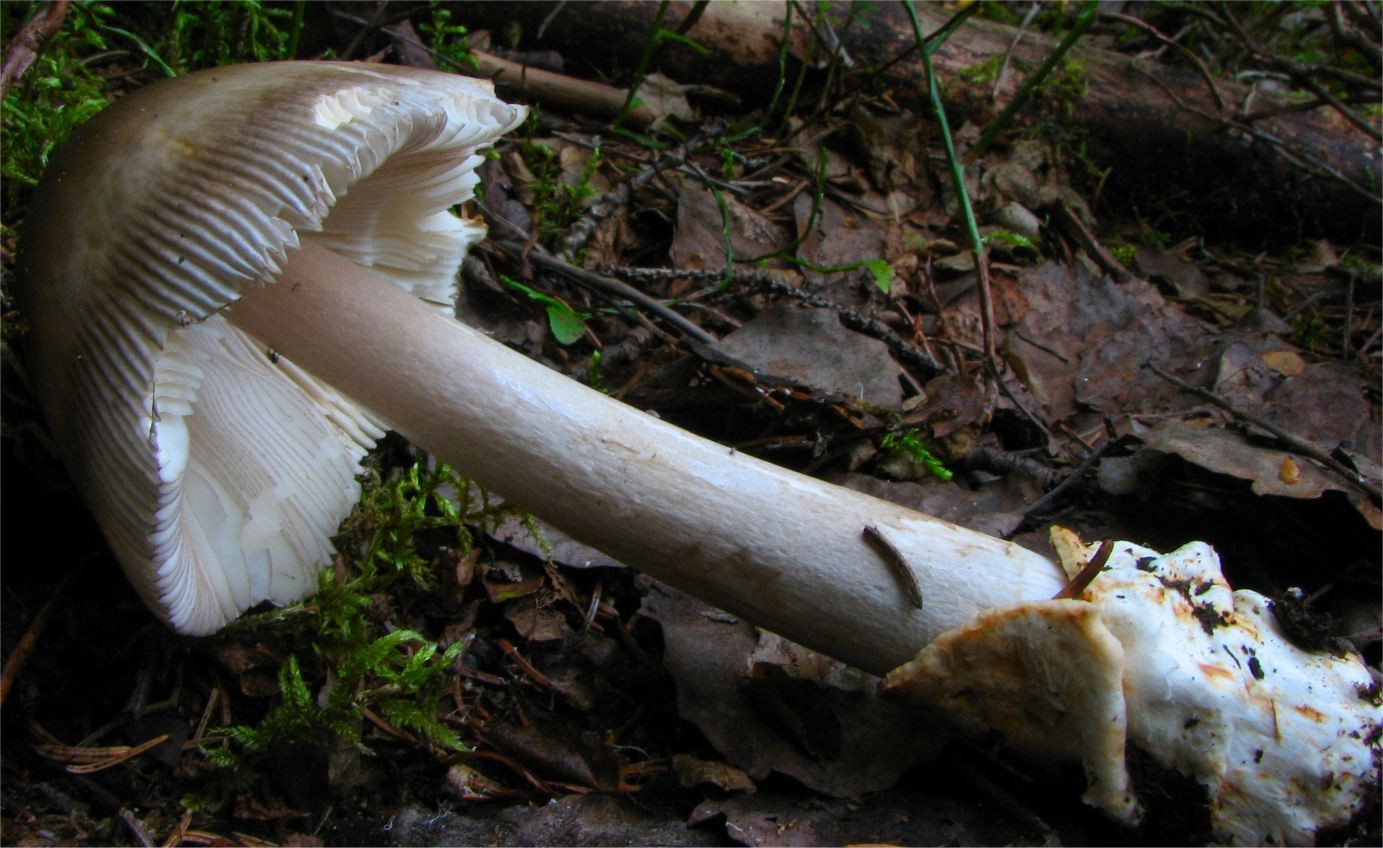
Irene Andersson (irenea) · CC BY-SA 3.0
Zmniejszono do maksymalnie 1600 pikseli i zapisano jako JPEG; zachowano proporcje, pełny kadr i barwy. Oryginał zachowano.
Photo source · CC BY-SA 3.0Amanita battarrae
Key differences
Muchomor brązowooliwkowy (Amanita submembranacea) forms caps more frequently covered with large veil patches, and its volva at the base of the stipe distinctly turns grey on the inner side.
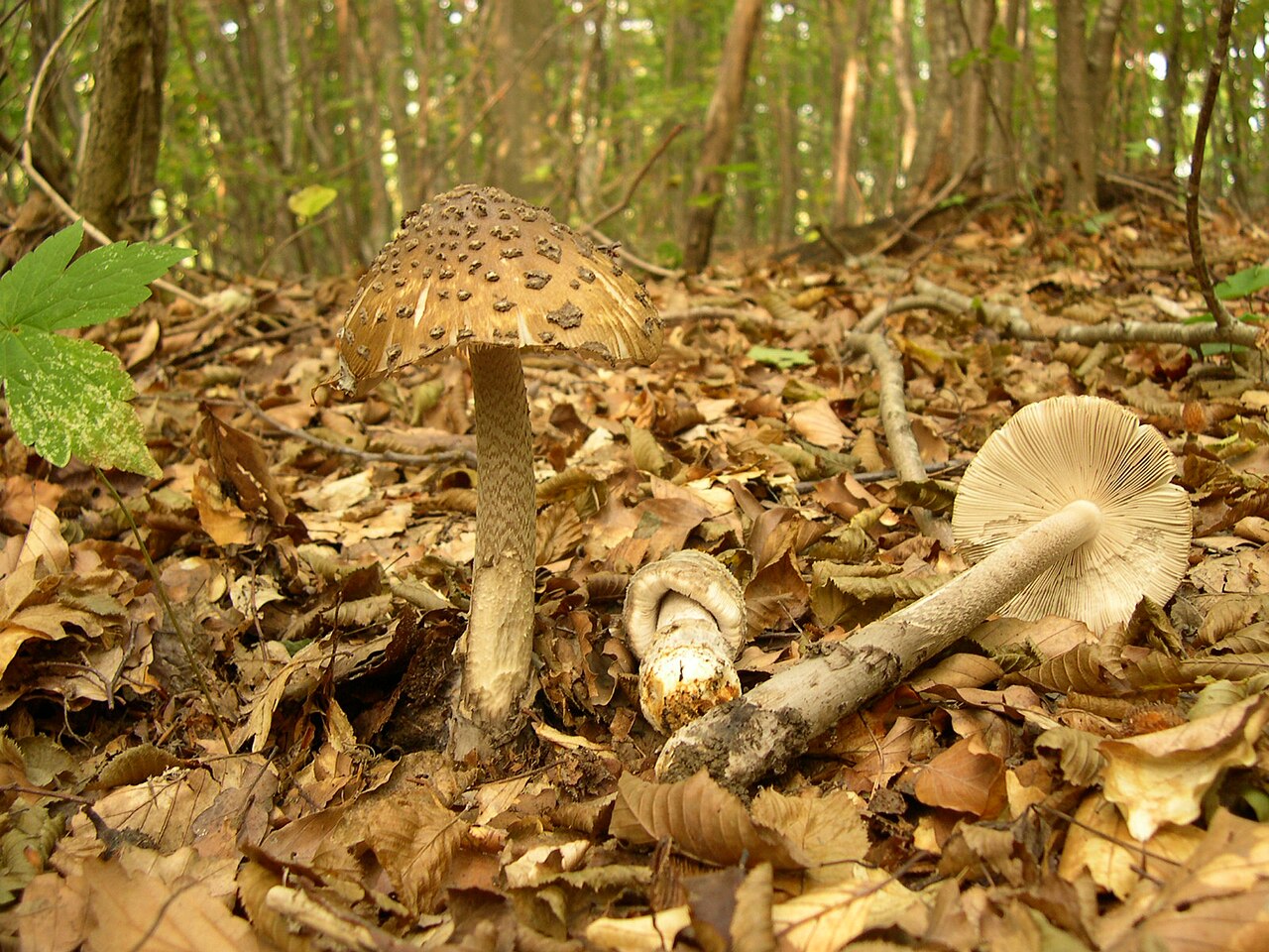
archenzo · CC BY-SA 3.0
Wersja JPEG o szerokości1280px przygotowana przez Wikimedia Commons; bez lokalnej zmiany kadru i koloru.
Photo source · CC BY-SA 3.0Amanita ceciliae
Key differences
Muchomor brązowooliwkowy (Amanita submembranacea) differs in having large veil remnants on the cap and an internally greying volva, growing mainly in mountain forests on acidic soils.
FAQ
Is Amanita submembranacea edible?
Yes, it is edible when thoroughly cooked, but it is toxic if eaten raw or undercooked.
Where does Amanita submembranacea primarily grow?
It grows primarily in montane and subalpine coniferous and mixed forests in European mountain ranges.
Which tree species are associated with Amanita submembranacea?
It associates mainly with conifers such as spruce, fir, larch, and pine, as well as mountain birch.
When is the fruiting period of this species?
It fruits during late summer and early autumn, typically from late July through September.
Why must Amanita submembranacea be thoroughly cooked?
Thorough heat treatment destroys the heat-labile hemolytic toxins that cause gastrointestinal illness.
Does Amanita submembranacea have a ring on its stem?
No, it belongs to Amanita section Vaginatae and lacks a partial veil and stem ring.
How does the volva of this species behave?
Its grayish submembranous volva is fragile and frequently breaks apart into crumbling fragments.
Is Amanita submembranacea used for non-culinary purposes?
Artisanal dyers use cap pigments with mordants to create earthy khaki and olive tones on wool.
In which regions is it traditionally eaten?
It is occasionally harvested for traditional household tables in alpine regions of Italy and Switzerland.
Was it used in educational collections?
Yes, casts and herbarium specimens are used in alpine forestry schools and museums for educational study.
Enthusiast
Names and history
- Obowiązująca nazwa krajowa
- muchomor brązowooliwkowy
- Obowiązująca nazwa taksonomiczna
- Amanita submembranacea (Bon) Gröger
- Inne nazwy krajowe
- Olive Amanita
- Pozostałe nazwy
- mglejarka brązowooliwkowa, gryzetka brązowooliwkowa
Home observations
Spore print
- Required materials
- fresh, mature cap, sheet of paper, glass or small bowl, water
- Time
- 2–24 h
- Procedure
- Place the cap underside down on the paper.
- Add a drop of water on top and cover.
- After 2–24 hours, lift the cap and inspect the deposit.
What to look for
- Spore print colour
- Spore print colour: white.
Under the microscope
- Required materials
- light microscope, microscope slide and cover slip, needle, water, spores from print
- Procedure
- Transfer a small amount of the spore print onto the slide using a needle.
- Add a drop of water, cover with a cover slip, and examine the spores.
What to look for
- Spore shape
- Spore shape: globose. Spore shape: subglobose. Spore shape: globose. Spore shape: globose. Spore shape: isodiametric (nearly, rarely).
- Spore surface
- Spore surface: smooth. Spore surface: smooth.
- Spore dimensions
- Length 9.5–13 µm, width 9–12 µm.
- Dimensions
- The rarely occurring spore form (nearly isodiametric shape) can have a length of 10 to 15 micrometres and a width of 9 to 13 micrometres.
Compare dimensions after calibrating the microscope scale.
Phenol experiment
- Material
- Basidiocarp context
- Reagent
- phenol
- Procedure
- In the described trial, phenol was applied to the indicated material.
- Conditions and source gaps
The source does not provide: sample preparation, phenol concentration, reagent amount, temperature, reading time, detailed method of reagent application.
Observation / result
- Source result
- In the flesh, after application of phenol, a dirty red-brown color was obtained.
- Source result
- In the flesh, after application of phenol, a wine-pink color was obtained.
Ecology and distribution
- Soil reaction
- Soil reaction: acidic.
- Soil fertility
- Soil fertility: fertile.
- Broadleaf trees in the vicinity
- Broadleaf trees in the vicinity: oak.
Broadleaf trees in the vicinity: beech.
Broadleaf trees in the vicinity: birch.
Broadleaf trees in the vicinity: alder.
Broadleaf trees in the vicinity: lime tree.
Broadleaf trees in the vicinity: hornbeam. - Coniferous trees in the vicinity
- Coniferous trees in the vicinity: spruce.
Coniferous trees in the vicinity: fir. - Shrubs in the vicinity
- Shrubs in the vicinity: Pinus mugo.
- Mycorrhizal hosts
- General species association: Mycorrhizal hosts: Abies (general partner list).
General species association: Mycorrhizal hosts: Betula (general partner list).
General species association: Mycorrhizal hosts: Larix (general partner list).
General species association: Mycorrhizal hosts: Picea (general partner list). - Mode of nutrition
- Mode of nutrition: ectomycorrhizal.
Non-culinary uses
Documented non-culinary applications of Amanita submembranacea are specialized, centered around botanical education, historical craft experimentation, and natural textile dyeing. In traditional artisan fiber work, textile dyers exploring mountain botanicals have documented the use of its dark olive-brown and bronze caps as a natural pigment source. When simmered with unspun wool or animal fibers and fixed with metallic mordants like iron or copper sulfates, the mushroom imparts muted khaki, olive-grey, and earthy brown tones, representing an interesting subject in historical natural dyeing circles focused on subalpine flora.
Beyond craft dyeing, Amanita submembranacea holds a defined non-culinary role in botanical archiving, academic collections, and environmental education. Dried herbarium exsiccata and historical plaster or resin casts of the species have been utilized by alpine forestry academies, national park visitor centers, and botanical museums across Central Europe. These physical models and preserved specimens serve to train forestry personnel, students, and mountain rangers in recognizing montane biodiversity and high-altitude fungal ecosystems without disturbing fragile alpine habitats.
History and culture
Amanita submembranacea was formally described in 1975 by French mycologist Marcel Bon (as Amanitopsis submembranacea) and transferred to Amanita by Frieder Gröger in 1979, and earlier documented in classical alpine surveys by mycologists examining high-altitude fungi. Historically, the species was often grouped within complex aggregates of the grisette group before being distinguished by its darker bronze-olive pigmentation and crumbling volva. Its cultural narrative is intimately bound to the cultural landscape of alpine Europe, where mountain communities developed nuanced vernacular traditions distinguishing high-altitude woodland fungi from valley varieties.
In alpine folklore, ringless amanitas growing near rocky treelines were often associated with local mountain legends and pastoral lore, viewed as quiet indicators of late-summer pasture transitions before the autumn cattle descent (Almabtrieb). Historical regional floras and mycological monographs from Swiss, French, and Italian alpine societies featured detailed lithographs of the species, documenting its place in the montane mycobiota. Unlike lowland mushrooms that entered urban literature and royal cuisine, Amanita submembranacea remained an emblem of rugged montane nature and provincial botanical exploration.
Edibility and safety
Amanita submembranacea is classified as an edible species following proper culinary preparation, but it contains raw toxins typical of ringless amanitas. Consuming the mushroom raw or undercooked results in gastrointestinal illness, stomach cramps, and hemolytic reactions. Rigorous and thorough thermal processing is an absolute prerequisite for safe ingestion; prolonged boiling, frying, or baking ensures complete destruction of all heat-labile harmful elements.
The cooked flesh is tender, delicate, and mild in flavor, though somewhat insubstantial owing to the thin-fleshed nature of the cap. Culinary utilization and consumer appreciation are primarily concentrated in alpine and subalpine communities where regional foragers have long gathered ringless amanitas. In mountain regions of northern Italy, Switzerland, and parts of Austria, it is occasionally harvested for traditional household tables. In non-mountainous regions and across the British Isles and North America, it is virtually unknown as a food source, and general culinary engagement is avoided due to widespread caution surrounding wild Amanita gathering.
Habitat and occurrence
Amanita submembranacea is a montane and subalpine species predominantly distributed across mountainous regions of Central, Southern, and Western Europe, with confirmed occurrences in the Alps, the Pyrenees, the Carpathians, the Apennines, and the Scandinavian mountains. It also occurs in montane sectors of Western Asia and temperate Eurasia. The species exhibits a strong ecological preference for high-elevation coniferous and mixed montane forests. Its primary tree associates are conifers such as Norway spruce (Picea abies), silver fir (Abies alba), European larch (Larix decidua), and Scots pine (Pinus sylvestris), though it also associates with mountain birch (Betula pubescens) and European beech (Fagus sylvatica) in upper montane belts.
The species colonizes acidic, humid, and rocky soils, frequently occurring among deep moss carpets, acidic mor humus, and mountain stream banks. Its fruiting season is comparatively short and closely tied to high-altitude summer weather. Sporocarps appear from mid-summer to early autumn, typically from late July through September. In lower-elevation montane valleys or sheltered ravines, fruiting can occasionally persist into mid-October depending on the onset of winter frosts.
Sources
- AMB-PESARO
- MYCOLOGIE-GRENOBLE
- NDFF Verspreidingsatlas: Amanita submembranacea
- Tulloss, Amanita Studies: Amanita submembranacea
- WOJ-2003
- Marek Snowarski/GRej:Amanita submembranacea
- NationalparkHarz, Großpilzflora, Band23
- src-kibby 2023
- UniversitéClaudeBernardLyon1, Champignons:Amanita submembranacea
- Marcel Bon 1975, protolog Amanitopsis submembranacea, Bull.Soc.linn.Lyon44(6): 176–179, DOI 10.3406/linly.1975.10188, s.178, OCR Persée
- Marek Snowarski, Grzyby.pl/GRej, Amanita submembranacea, dane obserwacyjne i bibliografia
- Wojewoda 2003, Checklist of Polish larger Basidiomycetes, s.50 (PDF52)
Researcher
Edibility and culinary use
Ustalenia źródłowe: jadalność, spożycie i wykorzystanie
- Required processing
- Requires preparation
Université Lyon1, Champignons: Amanita submembranacea, karta dydaktyczna · Comestibilité/Remarque: source condition cuisson; lack of a specific validated time and temperature protocol
- Edibility
- Edible — źródło oznaczenia
Materiał: po pełnej obróbce
Université Lyon1, Champignons: Amanita submembranacea, karta dydaktyczna · Comestible sous condition, Remarque; educational card, not a safety trial
- Raw consumption
- Poisonous
Materiał: surowy
Université Lyon1, Champignons: Amanita submembranacea, karta dydaktyczna · Remarque; Syndrome associé: source author links absence of cooking to harmful haemolytic effects. Does not establish a tested safe preparation protocol.
Scientific names and taxonomy
- Current scientific name
Amanita submembranacea (Bon) Gröger
Scientific names and taxonomy
Amanita submembranacea (Bon) Gröger belongs to the family Amanitaceae (Amanitaceae) and section Vaginatae. The current combination was published in 1979 based on the basionym Amanitopsis submembranacea Bon from 1975.
Scientific names and taxonomy
In historical mycological literature, the unpublished name Amanita subalpina M. M. Moser ined. has sometimes been associated with this taxon.
Taxon scope and identification limitations
A modern revision of the European section Vaginatae confirms the species distinctness of A. submembranacea based on a combination of microscopic volval characters with sequence analysis of nrDNA ITS, nrDNA LSU, RPB1, and TEF1-α.
Ecology and distribution
- Occurrence frequency
- Poland: Occurrence frequency: rare. [28]
Laboratory diagnostics
Dane ustrukturyzowane
Basidia
Other laboratory characteristics
- Apex of the gill edge cells
- Apex of the gill edge cells: capitate. [25]
- Constrictions of the gill edge cells
- Constrictions of the gill edge cells: constricted. [25]
- Course of the gill edge cells
- Course of the gill edge cells: tortuous. [25]
- Length of the gill edge cell
- Length of the gill edge cell: 20–40 µm. [37]
Length of the gill edge cell: 20–30 µm. [25] - Segmentation of the gill edge cells
- Segmentation of the gill edge cells: articulated. [25]
- Shape of other gill edge cells
- Shape of other gill edge cells: clavate (broad). [37]
Shape of other gill edge cells: spherical with a stalk (in some cells). [25]
Shape of other gill edge cells: shaped like an Opuntia segment. [25]
Shape of other gill edge cells: hammer-shaped. [25] - Regularity of the gill edge cell group
- Regularity of the gill edge cell group: irregular (often). [25]
- Width of the gill edge cell
- Width of the gill edge cell: 16–30 µm. [37]
Width of the gill edge cell: 10–15 µm. [25] - Gelatinization of the subhymenium
- Gelatinization of the subhymenium: gelatinized (slightly gelatinized, sometimes, at the gill edge). [25]
- Subhymenium structure
- Subhymenium structure: tortuous. [25]
Subhymenium structure: articulated (in the basidiophore zone). [25]
Subhymenium structure: subcellular (in the basidiophore zone). [25] - Cell types in the universal veil
- Cell types in the universal veil: sphaerocysts (partly). [25]
Cell types in the universal veil: filamentous hyphae. [25]
Cell types in the universal veil: filamentous hyphae (predominant, outer surface of the veil, at the stem base). [33]
Cell types in the universal veil: sphaerocysts (at the stem base, proportion increases, toward the interior). [33] - Diameter of spherical cells of the universal veil
- Diameter of spherical cells of the universal veil: 20–70 µm (sphaerocyst). [25]
- Shape of the universal veil cell
- Shape of the universal veil cell: globose. [25]
- Width of the universal veil hypha
- Width of the universal veil hypha: 3–15 µm. [25]
Pileipellis
- Arrangement of pileipellis hyphae
- Arrangement of pileipellis hyphae: Parallel. [31]
Arrangement of pileipellis hyphae: Parallel (hypocutis, more or less). [25] - Shape of pileipellis elements
- Shape of pileipellis elements: cylindrical. [37]
- Width of pileipellis elements
- Width of pileipellis elements: 10–15 µm (hypocutis). [25]
Width of pileipellis elements: 3–6 µm. [31]
Width of pileipellis elements: 3–6 µm (epicutis). [25] - Variability of pileipellis gelatinization
- Variability of pileipellis gelatinization: variable. [37]
Variability of pileipellis gelatinization: variable (epicutis). [25] - Embedding of cuticle hyphae in a gelatinous substance
- Embedding of cuticle hyphae in a gelatinous substance: present (variable degree of gelatinization, epicutis). [25]
Spores
- Spore colour under the microscope
- Spore colour under the microscope: hyaline. [31]
Spore colour under the microscope: yellow (rust-coloured, faint colour). [25] - Original assessment of the endosporium reaction in iodine
- Original assessment of the endosporium reaction in iodine: pseudoamyloid (weakly, result questioned in the source). [25]
- Position of the spore guttule
- Position of the spore guttule: central. [25]
- Droplets inside the spore
- Droplets inside the spore: present (large droplets). [25]
Hymenophoral trama
Material and research methods
Description of characteristics — source and scope
Material: General author description Lyon 1: Odeur faible, voire nulle.
Reading scope: Documented observation.
Limitations: Didactic description without specimen number; undetectable odor is a different aspect than weak intensity.
Description of characteristics — source and scope
Material: Studied Polish material in description Ronikier 2012, taste mild, odor none.
Reading scope: Documented observation.
Limitations: Author's description; not instructions for taste testing of raw mushroom.
Chemical reagent test — material and limitations
Material: Flesh of fruit bodies in description Bon 1975, paragraph Chair; phenol, without given concentration, temperature, or reading time.
Reading scope: Documented observation.
Limitations: Chronological color change and protocol from other species were not added. Original color term and custom qualifier remain separate.
Study of composition and activity — material and limitations
Material: Fruit bodies of A. submembranacea, intracellular silver; exact collective material described in the abstract.
Reading scope: Abstract.
Molecular study — Borovička et al. (2010)
Material: Silver complexes and their peptide ligand from fruit bodies of A. submembranacea; molecular separation of fractions in the abstract.
Scope of reading: Abstract.
Common limitations of source reading
Limitations: The entire abstract of the original publication was read via the EuropePMC index. The full article was not read; missing N and details of individual specimens. The aggregate range of 304–692 mg/kg for section Vaginatae was not assigned to this species. This is not evidence of bioremediation implementation or medicinal use. Methods listed in the abstract: size-exclusion chromatography, sulfhydryl fluorometry, RP-HPLC, and improved polyacrylamide gel electrophoresis. Unreported parameters were not supplemented. Species identification was provided by the authors (2010). The identification method and voucher specimen are not available in the read abstract; no redetermination of the material was performed.
Archiwalny opis siedliska
W Wielkiej Brytanii grzyb pojawia się w lasach liściastych i mieszanych, szczególnie pod dębami i bukami, z owocnikowaniem notowanym jesienią (m.in. w październiku).
Archiwalny opis siedliska
W Europie Zachodniej występuje na glebach kwaśnych w lasach liściastych i iglastych, zwłaszcza w rejonach podgórskich i górskich, w okresie od czerwca do listopada.
Archiwalny opis siedliska
W górach Harzu grzyb rośnie w lasach iglastych i mieszanych ze świerkiem na wysokości 280–1000 m n.p.m. (głównie powyżej 600 m), owocnikując od połowy czerwca do listopada.
Archiwalny opis siedliska
W Alpach włoskich owocniki zbierano w sierpniu w lasach z udziałem jodły i buka.
Archiwalny opis siedliska
Materiał potwierdzony badaniami genetycznymi dokumentuje występowanie gatunku w lasach Europy Środkowej i Zachodniej, w tym w Niemczech, Austrii, Belgii, Francji, Włoszech i Estonii.
- Results as recorded in the sources — Spores per basidium
- Liczba zarodników na jednej podstawce wynosi 4. [25] [37]
- Results as recorded in the sources — Spore diameter
- Materiał badania: Zbadane owocniki masywu Sarniej Skały 2000–2003, Polska; KRAM F-39947,F-50021,F-54314,F-54315,F-54316. Ryc. 9 wiąże struktury z KRAM F-39947. Ogólne metody: Nikon Eclipse E600, światło standardowe lub kontrast Nomarskiego; większość pomiarów w 5%KOH, czasem Melzer/CongoRed. 10–12 µm [37]
Materiał badania: Opis pierwotny Bon 1975: typ Fontanet koło LePuy, Francja,17.09.1974,nr 740917; dodatkowe zbiory 740912 i 740920 (s.179). Poszczególnych pomiarów zbiorczego opisu nie przypisano dowolnie jednemu okazowi. 11–13 µm (skrajnie 9–14 µm) [25]
Metody i zapisy źródłowe — Karta Société Mycologique du Dauphiné, opis i mikrofotografie R.G.; brak indywidualnego eksykatu dla opisu. Zdjęcia zarodników opisano jako w wodzie,400× i 1000×; ogólny opis hyalines nie ma osobno określonego medium.
Układ strzępek skórki — zapis źródłowy: „Cuticule ... +/- enchevêtrées ou parallèles; tu tylko alternatywa parallèles, bez przenoszenia +/- z określenia splątania.” [31]
Szerokość elementów skórki — zapis źródłowy: „Cuticule constituée d’hyphes allongées et +/- enchevêtrées ou parallèles, non bouclées, larges de 3 à 6 µm.” [31]
Barwa zarodnika w mikroskopie — zapis źródłowy: „hyalines” [31]
Metody i zapisy źródłowe — Opis pierwotny Bon 1975: typ Fontanet koło LePuy, Francja,17.09.1974,nr 740917; dodatkowe zbiory 740912 i 740920 (s.179). Poszczególnych pomiarów zbiorczego opisu nie przypisano dowolnie jednemu okazowi.
Identyfikacja kropli: nierozstrzygnięta.
Metoda źródłowa. Technika: mikroskopia świetlna (LM). Medium: iodine. Opis metody w źródle: Bon s.178 podaje jod, bez składu odczynnika, stężenia, czasu i osobnej kalibracji. Nie założono preparatu Melzera.
Układ strzępek skórki — zapis źródłowy: „Bon s.178: cellules sous-jacentes de l’hypocutis ... subparallèles.” [25]
Liczba zarodników na jednej podstawce — zapis źródłowy: „tétraspores” [25]
Długość podstawki; Szerokość podstawki — zapis źródłowy: „Basides55–70×15–20µm” [25]
Kształt podstawki — zapis źródłowy: „Basides allongées clavé-pédonculées; ogólna część kształtu maczugowatego. Wydłużenie i trzonkowatość zachowano oddzielnie w źródłowej dokumentacji.” [25]
Wierzchołek komórek ostrza blaszki — zapis źródłowy: „Bon s.178: capitées; wierzchołek główkowaty.” [25]
Przewężenia komórek ostrza blaszki — zapis źródłowy: „Bon s.178: étranglées; obecne przewężenia.” [25]
Przebieg komórek ostrza blaszki — zapis źródłowy: „Bon s.178: tortueuses; przebieg kręty.” [25]
Długość komórki ostrza blaszki; Szerokość komórki ostrza blaszki — zapis źródłowy: „Cellules marginales en partie sphéropédonculées (20–30 × 10–15 µm); pomiar tylko tej części komórek.” [25]
Członowanie komórek ostrza blaszki — zapis źródłowy: „Bon s.178: articulées; członowanie niezależne od kształtu.” [25]
Kształt innych komórek ostrza blaszki — zapis źródłowy: „Bon s.178: en partie sphéropédonculées; udział dotyczy wyłącznie tego kształtu.” [25]
Kształt innych komórek ostrza blaszki — zapis źródłowy: „Bon s.178: en raquettes d’Opuntia; odrębny źródłowy kształt, bez przeniesienia częstości nieregularności.” [25]
Kształt innych komórek ostrza blaszki — zapis źródłowy: „Bon s.178: et même en marteau; kształt młotkowaty.” [25]
Regularność grupy komórek ostrza blaszki — zapis źródłowy: „Bon s.178: mais souvent difformes; częsta nieregularność grupy komórek, bez przypisania jej arbitralnemu podstawowemu kształtowi.” [25]
Typ tramy hymenoforu — zapis źródłowy: „trame bilatérale” [25]
Szerokość miejscowego rozdęcia strzępki tramy — zapis źródłowy: „Bon s.178: avec parfois des hernies vers15–20µm; czasami miejscowe rozdęcia.” [25]
Szerokość strzępek tramy — zapis źródłowy: „Bon s.178: hyphes de toutes tailles×3–8µm; podstawowy zakres zwykłych strzępek.” [25]
Szerokość strzępek tramy — zapis źródłowy: „Bon s.178: jusque10–15µm pour les hyphes fondamentales.” [25]
Szerokość elementów skórki — zapis źródłowy: „Bon s.178: quelques extrémités libres égales ou clavées (×3–6µm).” [25]
Szerokość elementów skórki — zapis źródłowy: „Bon s.178: Hypocutis à hyphes épaissies vers10–15µm; wyłącznie hypocutis.” [25]
Zmienność zżelatynizowania skórki kapelusza — zapis źródłowy: „Bon s.178: plus ou moins gélifié; zmienność żelu w epicutis.” [25]
Osadzenie strzępek skórki w substancji żelowej — zapis źródłowy: „Epicutis plus ou moins gélifié en ixocutis; obecność żelu w epicutis. Nie wybrano niepodanego stopnia slight ani strong.” [25]
Organizacja skórki kapelusza — zapis źródłowy: „Bon s.178: Epicutis plus ou moins gélifié en ixocutis; kutis i obecność żelu, stopnia nie podano.” [25]
Barwa zarodnika w mikroskopie — zapis źródłowy: „Bon s.178: zone endosporique un peu jaune-roussâtre dans l’iode; barwa strefy endospory w jodzie.” [25]
Średnica zarodnika — zapis źródłowy: „Bon s.178: diamètre(9)–11–13–(14µm); typowe i skrajne granice osobno.” [25]
Źródłowa ocena reakcji endosporium w jodzie — zapis źródłowy: „Bon s.178: pseudoamyloïdité faible?; pytająca ocena autora, nie potwierdzony dodatni wynik.” [25]
Położenie kropli zarodnika — zapis źródłowy: „À grosse guttule centrale; centralne położenie opisanej kropli, oddzielnie od jej obecności i wielkości.” [25]
Krople wewnątrz zarodnika — zapis źródłowy: „Spores ... à grosse guttule centrale; obecność dużej guttuli, bez oznaczenia jej składu i bez dopisania liczebności z samego liczby pojedynczej słowa.” [25]
Długość zarodnika; Szerokość zarodnika — zapis źródłowy: „Bon s.178:10–15×9–13µm, wyłącznie rzadka forma subizodiametryczna.” [25]
Kształt zarodnika — zapis źródłowy: „Spores globuleuses” [25]
Kształt zarodnika — zapis źródłowy: „Bon s.178: rarement subisodiamétriques (par exemple10–15×9–13µm); rzadka forma niemal równowymiarowa.” [25]
Zżelatynizowanie podhymenium — zapis źródłowy: „Bon s.178: subgélifié parfois au niveau de l’arête; czasami nieznaczny żel przy ostrzu.” [25]
Budowa subhymenium — zapis źródłowy: „Bon s.178: Sous-hyménium tortueux.” [25]
Budowa subhymenium — zapis źródłowy: „Bon s.178: articulé puis subcelluleux dans la zone basidiophore; układ członowany tej strefy, nie chronologia wieku owocnika.” [25]
Budowa subhymenium — zapis źródłowy: „Bon s.178: subcelluleux dans la zone basidiophore; budowa subkomórkowa tej strefy.” [25]
Typy komórek w osłonie całkowitej — zapis źródłowy: „Bon s.178: en partie globuleuses; kuliste komórki stanowią część osłony, nie całość.” [25]
Typy komórek w osłonie całkowitej — zapis źródłowy: „Bon s.178: entrelacs d’hyphes filamenteuses; obecność nitkowatych strzępek osłony.” [25]
Średnica kulistych komórek osłony całkowitej — zapis źródłowy: „Bon s.178: hyphes du voile général en partie globuleuses (diamètre20–70µm).” [25]
Kształt komórki osłony całkowitej — zapis źródłowy: „Hyphes du voile général en partie globuleuses; zapis podstawowego kulistego kształtu. Udział i średnica 20–70 µm pozostają oddzielne.” [25]
Szerokość strzępki osłony całkowitej — zapis źródłowy: „Entrelacs d’hyphes filamenteuses (× 3–5–10–15 µm); pełne minimum i maksimum szerokości strzępek, nie średnica sferocyst.” [25]
Metody i zapisy źródłowe — Zbadane owocniki masywu Sarniej Skały 2000–2003, Polska; KRAM F-39947,F-50021,F-54314,F-54315,F-54316. Ryc. 9 wiąże struktury z KRAM F-39947. Ogólne metody: Nikon Eclipse E600, światło standardowe lub kontrast Nomarskiego; większość pomiarów w 5%KOH, czasem Melzer/CongoRed.
Liczba zarodników na jednej podstawce — zapis źródłowy: „4-spored” [37]
Długość podstawki; Szerokość podstawki — zapis źródłowy: „Basidia50–57×14–15µm” [37]
Długość komórki ostrza blaszki — zapis źródłowy: „Marginal cells 20–40 × 16–30 µm, broadly clavate; tylko pomiar długości, szerokość kształtu pozostaje osobną potrzebą.” [37]
Kształt innych komórek ostrza blaszki — zapis źródłowy: „Ronikier s.17: broadly clavate.” [37]
Szerokość komórki ostrza blaszki — zapis źródłowy: „Marginal cells 20–40 × 16–30 µm, broadly clavate; tylko pomiar szerokości.” [37]
Kształt elementów skórki kapelusza — zapis źródłowy: „Pileipellis made up of cylindrical hyphae” [37]
Zmienność zżelatynizowania skórki kapelusza — zapis źródłowy: „Ronikier s.17: (ixo)cutis; źródłowa zmienność oznaczenia żelatynizacji, bez zgadywania stopnia.” [37]
Organizacja skórki kapelusza — zapis źródłowy: „Ronikier s.17: an (ixo)cutis; podstawowa organizacja cutis.” [37]
Średnica zarodnika — zapis źródłowy: „Ronikier s.17: Spores10–12µm, globose; średnica, nie dwie osie.” [37]
Kształt zarodnika — zapis źródłowy: „globose” [37]
Powierzchnia zarodnika — zapis źródłowy: „smooth” [37]
Metody i zapisy źródłowe — src-kibby 2023
Metody i zapisy źródłowe — MYCOLOGIE-GRENOBLE
Metody i zapisy źródłowe — Mikroskopowa budowa pochwy w opisie Kibby (2023), s. 76; nie przypisano jej samowolnie jednej z kolekcji ilustrowanych fotografiami.
Typy komórek w osłonie całkowitej — zapis źródłowy: „Kibby s.76: predominantly filamentous on the outer surface.” [33]
Typy komórek w osłonie całkowitej — zapis źródłowy: „Kibby s.76: becoming progressively more swollen to form large sphaerocysts on the inner tissues; wzrost udziału sferocyst ku wnętrzu.” [33]
Names and taxon scope
Primary name: Muchomor brązowooliwkowy is the name proposed by Wojewoda in the Polish checklist of 2003, p. 50; a later Polish field report uses the same name. It is not a legally established name. The Ekologia.pl atlas also gives mglejarka brązowooliwkowa and gryzetka brązowooliwkowa; these have been retained as alternative names of atlas origin. No independent confirmation was found for the former labels szaropochwowy and mglejarka szaropochwowa.
Basionym and synonymy: Bon described Amanitopsis submembranacea in 1975, p. 176, based on material from France; type Fontanet,17.09.1974,no. 740917. The current combination in the registry is Amanita submembranacea (Bon) Gröger, Boletus 3 (2): 27 (1979). Species Fungorum also lists var. griseoargentata Contu 1986 as a synonym. This is the registry's opinion; the type of this variety was not examined. A. subalpina M. M. Moser ined. remains an unpublished historical name. Neither the former var. bispora, currently A. reidiana, nor the compared A. olivaceogrisea were included in the species data.
Molecular data: Varga et al. (2024), Table 1, report their own specimen DB-2018-07-31-1(AD25), Trafoi, South Tyrol, Italy, with sequences PP375774,PP375269,PP358527,PP273684. The remaining rows cover Estonia, Germany, Belgium, France, and Austria, and refer to earlier works. These entries were not treated as genetic verification of Polish specimens or as our own product.
Polish localities, habitat, and season
Herbarium material: Ronikier described basidiomata from the Sarnia Skała massif: KRAM F-39947 (20.06.2000),F-50021 (04.07.2000),F-54314 (03.07.2002),F-54315 (20.08.2002),F-54316 (16.08.2003), from an elevation of 910–1160 m, on soil, in beech forest, fir-spruce forest, and spruce forest. Fig. 9 was linked to F-39947. The report by Chachuła et al. documents a basidioma in 2018 near mountain pine,1665 m,11 October. These are published morphological identifications; no re-identification or DNA studies were performed here.
Atlas season and observations: The available Polish atlas Ekologia.pl indicates July or August–October. In the season row, the full range July–October has been retained with a reference to the atlas; this is a popular work without its own phenological curve. Early June collections from the Polish monograph and GRej remain separate observations. No typical season or peak was derived from them. The French June–November and British dates were not substituted for the Polish season.
Range and rarity: Wojewoda recorded Babia Góra,1370–1670 m, following Bujakiewicz 1993. Chachuła et al. (2019) document their own find and cite seven further regions of Poland. Their term rarely recorded was retained as an assessment from that work, not a modern measurement of abundance across the entire country. In GRej, a confirmed exsiccatum number was distinguished from a declaration of future material transfer. An entry in GRej itself does not constitute legal protection status.
Microscopic structure and examination methods
Polish material and methods: Ronikier's description is based on material from the study area deposited in KRAM and ZAMU. General methods included a Nikon SMZ 1500 stereomicroscope and a Nikon Eclipse E600, standard light or Nomarski contrast; most measurements were performed in 5% KOH, occasionally in Melzer's reagent or Congo red. The author does not indicate the medium for each measurement of this species nor the sample sizes. 5% KOH was not assumed as a condition for every number. Dimensions of structures were usually presented as length × maximum width.
Results and comparability: Ronikier reports globose and smooth spores, diameter 10–12 µm, absence of amyloidity, basidia 50–57 × 14–15 µm with 4 spores. Bon describes basidia 55–70 × 15–20 µm with 4 spores and spore diameter (9)–11–13–(14) µm; rare forms measure 10–15 × 9–13 µm. Kibby gives 9,5–13 × 9–12 µm. Diameter was not transcribed as two axes, and rare forms from Bon were not added to an unconditional combined measurement. Source ranges and extreme limits remain separated.
Gill edge cells and volva: In the Polish material, gill edge cells measure 20–40 × 16–30 µm and are broadly clavate. Bon describes some cells as sphaeropedunculate 20–30 × 10–15 µm, and the remaining ones as distorted, articulate, flexuous, constricted, capitate, or hammered. They were not automatically equated with cystidia. Bon's veil contains loosely connected globose cells 20–70 µm and intertwined hyphae 3–15 µm. In Kibby, the outer side of the volva is predominantly filamentous, becoming progressively more swollen toward the interior to form sphaerocysts. Different layers and materials were not merged into a single unconditional result.
Pileipellis, trama, and clamps: Ronikier defines the pileipellis as an (ixo)cutis of cylindrical hyphae and reports the absence of clamps. Bon describes a gelatinized epicutis with free ends that are cylindrical or clavate, width 3–6 µm, and a lower layer with a different arrangement and pigment; hypocutis has hyphae 10–15 µm, among narrower ones 3–7 µm. Lamellar trama is bilateral, distinguishing regular hyphae, basal elements, and inflations. The general absence of clamps was not turned into an implicit examination of every cell base. Grenoble reports hyaline spores; the medium for this general assessment remains unspecified, separate from photo captions in water.
Chemical reactions: Bon describes a dirty reddish-brown to vinaceous-pink color with phenol, distinguishing it from an intense blackcurrant color. He describes the spores as inamyloid, although in iodine he notes a yellowish-rufous endospore zone and questions weak pseudoamyloidity. The question mark was not treated as a positive result nor equated with classic amyloidity of the entire spore. For the negative result in the Polish material, reagent concentration, temperature, and duration were not guessed.
Edibility and strength of toxicological evidence
Authors' assessments: Grzyby.pl provides an edible assessment, and the Lyon educational card 1 — conditionally edible. Lyon attributes haemolysins, haemolytic symptoms, and the need for cooking to the Amanitopsis group, but does not provide a specific toxin study of this species or a description of a specific patient series to support this thesis. This is not a validated safety protocol A. submembranacea. The distinction between the author's assessment, the warning for the group, and the species study result has been preserved.
ANSES report and limitations: ANSES compiled French poison reporting from 2017, excluding only cases with zero link to exposure. The report mentions A. phalloides and A. submembranacea as taxa identified from photographs among four cases of severe poisoning presenting with an amatoxin profile. It does not divide the number of cases between the two species, nor does it provide individual documentation, fruitbody toxin analyses, or data excluding a mixed meal. It does not permit attributing all 25 cases or two deaths to this species, nor proving the presence of amatoxins in its correctly identified fruitbodies. The mention was not omitted, but it was not given greater causal weight than documented in the publication.
Review status: In the examined material, no specific analysis of haemolysins or amatoxins for this species was found, nor full individual clinical documentation that would resolve the above discrepancies. This is a limitation of the review, not proof of the absence of toxins or studies. The entry edible after thorough cooking was designated as an assessment by the author of the educational card. It was not converted into a clinical outcome or into a deadly poisonous category.
Silver accumulation and other uses
Species study result: Borovička et al. (2010) analyzed silver complexes in fruitbodies of A. submembranacea using partition chromatography, thiol ligand assays, and electrophoresis. The entire abstract reviewed states that nearly all analyzed silver was intracellular, predominantly in 7 kDa complexes and in a smaller 3,3 kDa fraction; in isolated complexes, a single thiol peptide of approximately 3 kDa was detected. This suggests the involvement of metallothionein, not a complete identification of the mechanism in every specimen.
Reading level and scope: The complete abstract of the primary publication was reviewed; the full text with tables, specimen numbers, and methods was not obtained. The range 304–692 mg/kg applies in the abstract to Amanita spp. section Vaginatae, so its boundaries were not attributed to a single species. The absence of glutathione and phytochelatins refers to the isolated complexes studied, without asserting their biological absence in the entire species. The results of the experiment by Osobová et al. (2011) on A. strobiliformis were not transferred here.
Application vs. potential: The study documents metal accumulation and binding. It does not confirm the implementation of this species in bioremediation, pharmaceutical production, supplements, or silver extraction. Separate searches for technical, herbal, medicinal, dye, and culinary spice uses did not yield a primary source confirming such a practice within the reviewed scope. This does not imply proof of its nonexistence. Missing applications were not replaced with applications of other Amanita species.
Source documentation
Source findings: Tulloss lists trees Abies, Betula, Larix, and Picea for Amanita submembranacea, including examples of Picea abies, Abies alba, and Larix decidua. He points out that some collections from England, Norway, and Scotland formerly identified as A. submembranacea turned out to be A. reidiana. This is a qualification regarding the identification of these collections; it does not confirm their occurrence as A. submembranacea sensu stricto.
Occurrence verification — scope and limitations
Material: Polskie okazy KRAM F-39947,F-50021,F-54314,F-54315,F-54316 z masywu Sarniej Skały; stanowisko Babiogórskiego PN 2018 i polskie rekordy GRej.
Scope of review: Documented observation.
Review date: 18.10.2026.
Limitations: Without independent re-identification or genotyping. Collection dates do not establish season boundaries.
Occurrence verification — scope and limitations
Material: Zbiór z województwa małopolskiego oznaczony przez Błażeja Gierczyka.
Reading scope: Source fragments.
A source fragment was read; full text and methods were not verified.
Review date: 21.10.2026.
Limitations: Source identification and conceptual scope retained. No new examination of specimens or reading of all publications cited by the source was declared.
Scope of elaboration
Required checks are settled.
Legal basis and scope of assessment
Analytical results
Composition and laboratory activity
- Result of component detection in material
- Result of component detection in material: detected (Ag, fruit body preparation, identified in the source, intracellularly). [7]
- Molecular mass of the metal-binding ligand
- Molecular mass of the metal-binding ligand: 3 kDa (Ag, thiol peptide, hypothesized identity, hypothesis: metallothionein, silver-binding ligand, reported value). [7]
- Molecular mass of the metal complex
- Molecular mass of the metal complex: 7 kDa (Ag, metal complex, intracellularly, major component, major fraction of the silver complex, reported value). [7]
Molecular mass of the metal complex: 3.3 kDa (Ag, metal complex, intracellularly, minor component, minor fraction of the silver complex, reported value). [7]
Key substances and their effects
Key substances and their effects
In fruit bodies from sites with elevated metal content, intracellular binding and retention of silver were investigated; this element is bound mainly in peptide complexes of 7 kDa and 3,3 kDa, likely involving metallothioneins.
Key substances and their effects
In amanitas from section Vaginatae from polluted areas, silver concentrations reaching 304–692 mg/kg have been recorded.
Key substances and their effects
Consumption of raw amanitas from this group causes hemolytic poisoning manifested by nausea, vomiting, breakdown of red blood cells, hematuria, and the risk of kidney damage.
Ecological studies
Source documentation
Source findings: NDFF describes the only Dutch find of Amanita submembranacea under hornbeam in an avenue, on nutrient-rich soil. Outside the Netherlands, it lists sites under coniferous and deciduous trees, especially on acidic soils. The description of a single Dutch site was kept separate from the broader ecology; occurrence under hornbeam does not become evidence of mycorrhiza with this tree nor a general preference for fertile soils.
Habitat and occurrence
Potwierdzony zasięg występowania
Wszystkie obszary potwierdzone w zapisanych danych źródłowych.
| Kraj | Regiony i stanowiska | Fruiting | Sources |
|---|---|---|---|
| Poland | Lesser Poland Voivodeship | VII–X |
|
Sources
- Société Mycologique du Dauphiné, Amanita submembranacea, opis i mikrofotografie R.G. (także [31])
- NDFF Verspreidingsatlas: Amanita submembranacea
- Tulloss, Amanita Studies: Amanita submembranacea
- WOJ-2003
- Borovička i in. 2010 — wyniki abstraktu, odczyt 2026-10-19
- Minister Środowiska, Dz.U. 2014 poz. 1408, rozporządzenie w sprawie ochrony gatunkowej grzybów
- Minister Zdrowia, Dz.U. 2026 poz. 258, tekst jednolity rozporządzenia dotyczącego grzybów dopuszczonych do obrotu
- Marek Snowarski/GRej:Amanita submembranacea
- src-kibby 2023 (także [33])
- NCBITaxonomy619305
- Borovička i in. 2010, Science of the Total Environment408:2733–2744, DOI 10.1016/j.scitotenv.2010.02.031; pełny abstrakt w EuropePMC (także [27])
- Chloé Greillet/ANSES 2018, Twice as many poisonings and serious cases due to mushrooms in 2017 as in 2016, Vigil’Anses6:1–3
- Marcel Bon 1975, protolog Amanitopsis submembranacea, Bull.Soc.linn.Lyon44(6): 176–179, DOI 10.3406/linly.1975.10188, s.176, OCR Persée
- Marcel Bon 1975, protolog Amanitopsis submembranacea, Bull.Soc.linn.Lyon44(6): 176–179, DOI 10.3406/linly.1975.10188, s.178, OCR Persée
- Marcel Bon 1975, protolog Amanitopsis submembranacea, Bull.Soc.linn.Lyon44(6): 176–179, DOI 10.3406/linly.1975.10188, s.179, OCR Persée
- Chachuła i in. 2019, Grzyby wielkoowocnikowe stwierdzone w2018r. w Babiogórskim PN, Przegląd Przyrodniczy30(3): 3–26
- Ekologia.pl, Muchomor brązowooliwkowy — atlas, nazwy zwyczajowe i sezon polski
- Marek Snowarski, Grzyby.pl/GRej, Amanita submembranacea, dane obserwacyjne i bibliografia
- Species Fungorum, rekord 308592, synonimia Amanita submembranacea
- Muchomor brązowooliwkowy — polskie wykazy ochrony i obrotu
- Université Lyon1, Champignons: Amanita submembranacea, karta dydaktyczna
- API ELI Sejmu — bieżący status Dz.U.2014poz.1408
- Anna Ronikier 2012, Fungi of the Sarnia Skała massif in the Tatra Mountains, Polish Botanical Studies28, s.12–13 i16–17
- API ELI Sejmu — bieżący status Dz.U.2026poz.258
- Varga i in. 2024, Phylogenetic and morphological studies reveal large diversity and three new species in Amanita sect.Vaginatae from Europe, Mycological Progress 23, article 38, DOI 10.1007/s11557-024-01974-0
- Wojewoda 2003, Checklist of Polish larger Basidiomycetes, s.50 (PDF52)