
Edible
Requires preparation

Poisonous when raw
Set your location to see data for your region.


Amanita battarrae, commonly known as the banded grisette, is an ectomycorrhizal agaric characterized by a distinctly striate cap margin, concentric brownish-olive zonal banding, fragile flesh, and the complete absence of a persistent partial veil ring. Belonging to Amanita section Vaginatae, it produces tall, slender basidiocarps arising from an elongated, sheath-like white to rusty-stained volva nestled in forest litter, predominantly in temperate and boreo-alpine woodlands.
Identification, habitat, and key differences.
Forager
Fruitbody structure
- Fruit body form
- Fruit body form: Cap, stipe.
- Fruitbody height
- Fruitbody height: 150 mm.
Cap
- Cap shape
- in young fruit bodies: Cap shape: ovoid.
Cap shape: bell-shaped.
Cap shape: convex.
in mature fruit bodies: Cap shape: flat.
in young fruit bodies: Cap shape: hemispherical. - Cap surface relief
- Cap surface relief: smooth.
- Cap covering
- Cap covering: glabrous.
- Cap surface sheen
- Cap surface sheen: shiny.
- Cap stickiness and sliminess
- Cap stickiness and sliminess: sticky.
- Cap colour
- Cap colour: gray.
Cap colour: grey-brown.
Cap colour: yellow-olive. - Cap margin outline
- Cap margin outline: even.
- Cap margin surface relief
- Cap margin surface relief: grooved.
- Cracking and splitting of the margin
- Cracking and splitting of the margin: uncracked.
- Cap diameter
- The cap measures 40–120 mm in diameter.
- Cap centre
- Cap centre: with a distinct umbo.
Cap centre: with a distinct umbo.
Cap centre: Depressed. - Peelability of the cap cuticle
- Peelability of the cap cuticle: partially peelable.
Peelability of the cap cuticle: partially peelable (up to half). - Presence of veil remnants on the cap
- Presence of veil remnants on the cap: absent.
- Cap colour zoning
- Cap colour zoning: concentric (distinct zones).
Cap colour zoning: concentric.
Cap underside
- Hymenophore type
- Hymenophore type: gills.
Gills at stipe
- Hymenophore attachment to the stipe
- Hymenophore attachment to the stipe: free from the stipe (gills).
Hymenophore attachment to the stipe: attached to the stipe (gills).
Hymenophore attachment to the stipe: free from the stipe (gills).
Hymenophore attachment to the stipe: attached to the stipe (gills).
Hymenophore attachment to the stipe: free from the stipe (gills).
- Hymenophore colour
- Hymenophore colour: cream.
Hymenophore colour: whitish.
Hymenophore colour: creamy white.
Hymenophore colour: white. - Macroscopic structure of the free gill edge
- Macroscopic structure of the free gill edge: entire, with a smooth outline.
Macroscopic structure of the free gill edge: floccose.
in young fruit bodies: Macroscopic structure of the free gill edge: floccose.
in old fruit bodies: Macroscopic structure of the free gill edge: toothed or serrate. - Natural colour of the gill edge
- Natural colour of the gill edge: white.
- Gill spacing
- Gill spacing: closely spaced.
Gill spacing: widely spaced.
Gill spacing: rather widely spaced.
Gill spacing: closely spaced.
Gill spacing: crowded. - Gill thickness
- Gill thickness: thick.
- Gill depth
- Gill depth: up to 7 mm.
- Gill width class
- Gill width class: broad.
- Presence of short gills
- Presence of short gills: present.
Stem and base
- Stipe position
- Stipe position: central.
- Stipe shape
- Stipe shape: cylindrical.
Stipe shape: tapering upwards.
Stipe shape: club-shaped. - Stipe colour
- in the upper part of the stipe: Stipe colour: whitish.
Stipe colour: white. - Stipe surface relief
- in the upper part of the stipe: Stipe surface relief: grooved.
- Stipe surface covering
- Stipe surface covering: scaly.
- Colour of stipe ornamentation
- Colour of stipe ornamentation: grey-brown.
- Stipe interior
- Stipe interior: solid.
Stipe interior: hollow. - Stipe length
- Stipe length: 50–200 mm.
- Stipe diameter
- Stipe diameter: 5–20 mm.
- Volva
- Volva: present.
Volva: present.
Volva: present.
Volva: present.
Volva: present.
Ring
- Ring presence
- Ring presence: no ring.
Ring presence: no ring.
Ring presence: no ring.
Ring presence: no ring.
Universal veil at base
- Veil structure at the stipe base
- Veil structure at the stipe base: sac-shaped.
Veil structure at the stipe base: sac-shaped.
Veil structure at the stipe base: sac-shaped.
- Color of volva and veil at stem base
- Color of volva and veil at stem base: white (veil structure at the stem base: volva).
Color of volva and veil at stem base: white (veil structure at the stem base: volva).
Color of volva and veil at stem base: white (veil structure at the stem base: volva).
Color of volva and veil at stem base: white (veil structure at the stem base: volva). - Stem robustness
- Stem robustness: stout.
- Material of volva and veil at stem base
- Material of volva and veil at stem base: membranous (veil structure at the stem base: volva).
Flesh
- Flesh colour
- Flesh colour: white.
Flesh colour: whitish (stipe flesh). - Flesh taste
- Flesh taste: astringent.
Flesh taste: weak. - Flesh resistance to breaking or tearing
- Flesh resistance to breaking or tearing: brittle.
- Flesh consistency type
- Flesh consistency type: fleshy (cap flesh).
- Flesh colour after damage
- Flesh colour after damage: no colour change.
- Flesh odour
- Flesh odour: pleasant (flesh).
Where and when it grows
- Habitat
- Habitat: coniferous woodland.
Habitat: mixed woodland.
Habitat: deciduous woodland. - Substrate
- Substrate: soil.
Substrate: litter. - Fruit body growth pattern
- Fruit body growth pattern: solitary.
Fruit body growth pattern: scattered groups.
Fruit body growth pattern: in small groups. - Fruiting season
- Fruit bodies appear from June to September.
- Distribution area
- Distribution area: confirmed.
Edibility and culinary use
- Edibility
- Edible
- Required processing
- Requires preparation
- Raw consumption
- Poisonous when raw
Law
- Protection
- It is not subject to species protection in Poland.
- Trade
- It is approved in Poland for neither trade nor the production of mushroom products.
Possible confusions
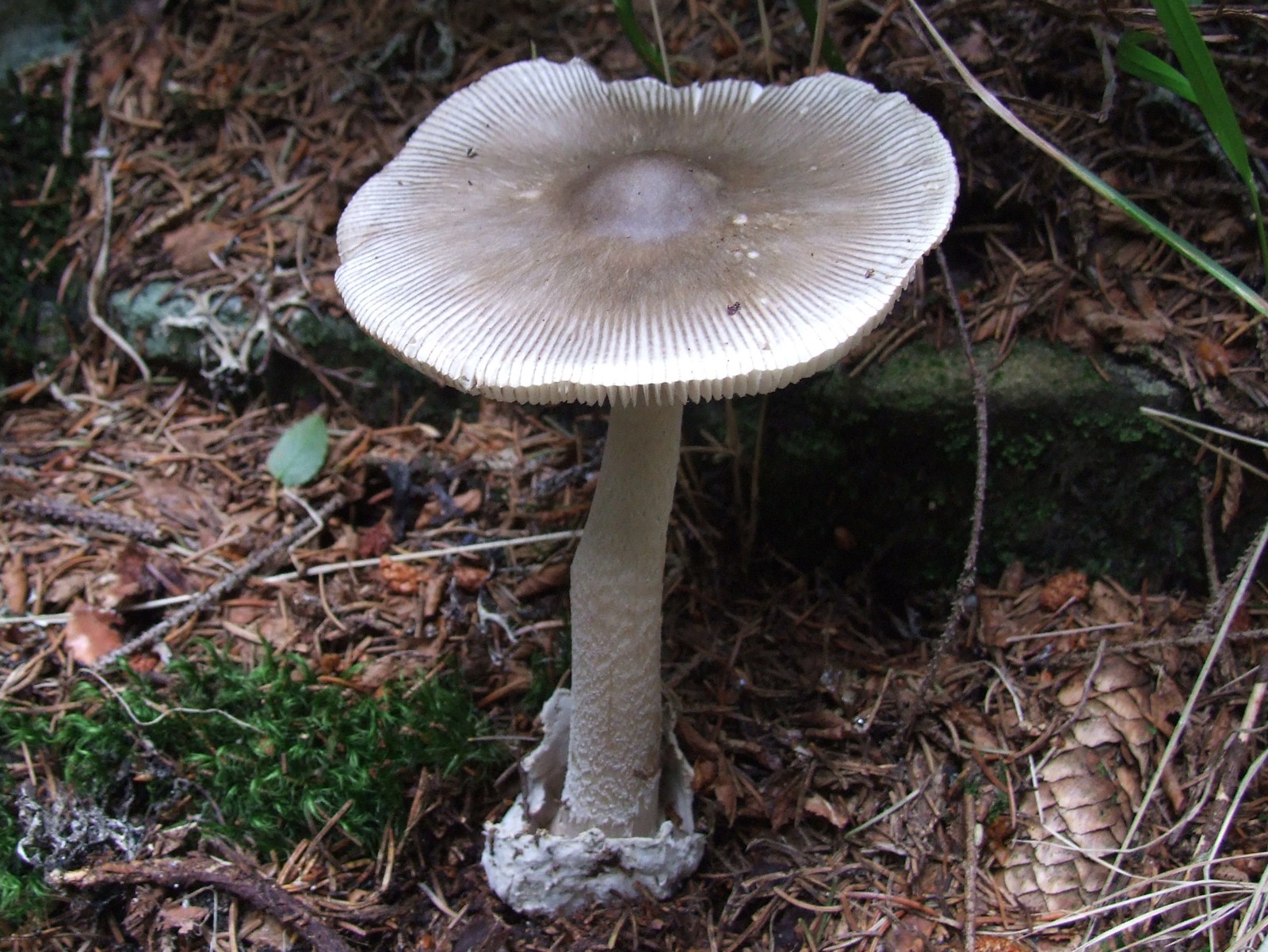
Jerzy Opioła · CC BY-SA 4.0
Zmniejszono do maksymalnie 1600 pikseli i zapisano jako JPEG; zachowano proporcje, pełny kadr i barwy. Oryginalny plik źródłowy zachowano.
Photo source · CC BY-SA 4.0Amanita vaginata
Key differences
The grey amanita (Amanita vaginata) differs from the olive amanita by a cap of uniform shade without concentric zonation and a smooth, white stipe lacking darker scales.
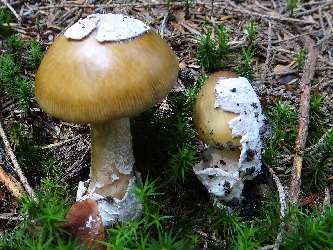
Jerzy Opioła · CC BY-SA 3.0
Wersja JPEG o szerokości 1280 px przygotowana przez Wikimedia Commons; bez dodatkowych lokalnych zmian.
Photo source · CC BY-SA 3.0Amanita submembranacea
Key differences
Muchomor brązowooliwkowy (Amanita submembranacea) forms caps more frequently covered with large veil patches, and its volva at the base of the stipe distinctly turns grey on the inner side.
Amanita fulva
Key differences
Tawny grisette (Amanita fulva) is distinguished by the warm, fulvous-orange or fox-brown color of the pileus, white lamellae with an unchanging edge, and a smooth stipe.
FAQ
Does Amanita battarrae have a ring on its stem?
No, it belongs to the ringless group of amanitas and lacks any annulus or stem collar.
Can Amanita battarrae be eaten raw?
No, it contains heat-labile toxins that trigger severe stomach illness if consumed raw.
What primary trees does this species associate with?
It forms mycorrhizae primarily with spruce, fir, pine, beech, birch, and chestnut.
In what months is it most commonly observed?
Fruiting typically extends from July through October depending on regional elevation.
Where does the name battarrae come from?
It honors Giovanni Antonio Battarra, an eighteenth-century Italian naturalist and priest.
Can wool be dyed using this mushroom?
Yes, with metal mordants it produces soft beige, pale tan, and muted olive tones on wool.
What type of soil does this species prefer?
It favors acidic to neutral, well-drained soils rich in humus and leaf or needle litter.
Is this species popular for consumption in Western Europe?
No, Western European foraging guides generally discourage its consumption.
Does the cap have noticeable margin markings?
Yes, it displays distinctly grooved, combed striations along the entire cap margin.
Is the universal veil visible on the stem base?
Yes, the base of the stem is enclosed in an elongated, sack-like, tall white volva.
Enthusiast
Names and history
- Obowiązująca nazwa taksonomiczna
- Amanita battarrae
- Inne nazwy krajowe
- Banded Amanita
- Pozostałe nazwy
- Grey-zoned Ringless Amanita, muchomor wielkopochwowy
Home observations
Spore print
- Required materials
- fresh, mature cap, sheet of paper, glass or small bowl, water
- Time
- 2–24 h
- Procedure
- Place the cap underside down on the paper.
- Add a drop of water on top and cover.
- After 2–24 hours, lift the cap and inspect the deposit.
What to look for
- Spore print colour
- Spore print colour: white.
Under the microscope
- Required materials
- light microscope, microscope slide and cover slip, needle, water, spores from print
- Procedure
- Transfer a small amount of the spore print onto the slide using a needle.
- Add a drop of water, cover with a cover slip, and examine the spores.
What to look for
- Spore shape
- Spore shape: globose. Spore shape: globose. Spore shape: globose. Spore shape: globose. Spore shape: subglobose. Spore shape: ellipsoid (broad). Spore shape: globose (nearly). Spore shape: ellipsoid (broad). Spore shape: ellipsoid (occasionally). Spore shape: globose (occasionally).
- Spore surface
- Spore surface: smooth. Spore surface: smooth. Spore surface: smooth. Spore surface: smooth.
- Spore dimensions
- Length 9–17.7 µm, width 7.2–15.6 µm.
Compare dimensions after calibrating the microscope scale.
Phenol experiment
- Material
- Basidiocarp context
- Reagent
- phenol
- Procedure
- In the described trial, phenol was applied to the indicated material.
- Conditions and source gaps
The source does not provide: sample preparation, phenol concentration, reagent amount, temperature, reading time, detailed method of reagent application.
Observation / result
- Source result
- In the context after applying phenol, the color described in the source as "violet" was obtained.
- Source result
- In the context after applying phenol, the color described in the source as "vinaceous red-brown" was obtained.
Ecology and distribution
- Soil reaction
- Soil reaction: acidic.
- Broadleaf trees in the vicinity
- Broadleaf trees in the vicinity: birch.
Broadleaf trees in the vicinity: oak.
Broadleaf trees in the vicinity: beech.
Broadleaf trees in the vicinity: hornbeam.
Broadleaf trees in the vicinity: lime tree. - Coniferous trees in the vicinity
- Coniferous trees in the vicinity: spruce.
Coniferous trees in the vicinity: fir.
Coniferous trees in the vicinity: pine. - Mode of nutrition
- Mode of nutrition: ectomycorrhizal.
Non-culinary uses
Amanita battarrae has very limited non-culinary utility, though it possesses documented historical relevance in natural history documentation, mycology education, and artisan fabric dyeing. Natural dye artisans have occasionally utilized mature basidiocarps of grisettes with iron and alum mordants, yielding soft tan, muted olive-beige, and pale gray-brown hues on unbleached sheep wool and animal fibers, although colorfastness is modest compared to polypores.
Historically, nineteenth- and twentieth-century botanical academies employed dried exsiccata and plaster or wax cast models of this species as morphological teaching tools to demonstrate the structural diversity of the genus Amanita and the anatomy of universal volval remnants. Furthermore, dried herbarium collections serve as reference standards in regional biodiversity mapping, and baseline spore deposits have historically been utilized in educational microscopy demonstrations illustrating unornamented, inamyloid basidiospore characteristics in classical taxonomy.
History and culture
Amanita battarrae owes its specific epithet to Father Giovanni Antonio Battarra (1714–1789), an Italian naturalist, mycologist, and priest from Rimini whose 1755 illustrated work 'Fungorum Agri Ariminensis Historia' documented numerous regional agarics. The basionym was published by Boudier in 1902 as a variety, raised to species rank by Riel in 1907, and subsequently transferred into modern taxonomy by Marcel Bon in 1985, standing as a tribute to eighteenth-century natural philosophy.
In European folk traditions, ringless amanitas or grisettes were rarely distinguished at the species level by rural gatherers, being grouped under dialectal names meaning 'gray caps' or 'sheathed fungi.' Because of their fragile nature and ominous botanical family, they often acquired an aura of ambivalence in local folklore. In Italian and French rural herbals, they were occasionally depicted in natural history chronicles as emblems of fleeting woodland beauty, admired for their delicate concentric zoning yet approached with caution by village herbalists who respected the unpredictable nature of wild forest agarics.
Edibility and safety
Amanita battarrae is considered conditionally edible after thorough and prolonged thermal processing, as raw specimens contain heat-labile hemolytic compounds that disrupt red blood cells and provoke acute gastroenteritis. Thorough boiling or sustained frying is strictly required before any consumption to denature these thermolabile toxins.
Even when cooked, consumption is discouraged in many regions due to personal sensitivities and the high risk of severe digestive distress. Culinary appreciation varies across national traditions: in parts of Eastern Europe and central European rural areas, experienced foragers traditionally cook grisettes thoroughly and consume them, regarding their flavor as mild, nutty, or somewhat sweet. Conversely, culinary guides across Western and Northern Europe widely classify the species as unsuitable or inadvisable for table use, emphasizing that individual gastrointestinal tolerance fluctuates significantly and that consumption offers modest culinary reward relative to the preparation demands.
Habitat and occurrence
Amanita battarrae occurs across temperate, montane, and boreo-alpine zones of Europe and parts of northern Asia, with related lineages documented across cooler parts of North America. It establishes ectomycorrhizal relationships with both deciduous and coniferous trees. Primary broadleaf hosts include European beech (Fagus sylvatica), birch (Betula), hornbeam (Carpinus betulus), and sweet chestnut (Castanea sativa), while in upland and northern regions it associates heavily with Norway spruce (Picea abies), fir (Abies alba), and scots pine (Pinus sylvestris).
The species prefers well-drained, acidic to neutral soils rich in organic matter, sandy loams, and moss-carpeted forest floors along mature woodland borders. Fruiting typically spans from mid-summer through late autumn, peaking between August and October in lowland central Europe. In subalpine or northern boreal regions, fruiting begins as early as July and concludes by September before the onset of autumn frosts, reflecting marked sensitivity to ambient ground temperature and seasonal precipitation.
Sources
- FIRST-NATURE
- Karel Tejkal/Česká mykologická společnost, Amanita battarrae
- GruppoMicologicoCastellano, Scheda0121
- Marek Snowarski/GRej, Amanita battarrae
- Kumar J., Atri N.S. 2020, Kavaka54:30–37, DOI 10.36460/Kavaka/54/2020/30-37
- Aalto M. 1974, Amanita magnivolvata sp.nova, Karstenia14:93–96; DOI 10.29203/ka.1974.92
- Tervonen K., Oldén A., Halme P. 2019. Ectomycorrhizal fungi in wood-pastures: Communities are determined by trees and soil properties, not by grazing. Agriculture, Ecosystems & Environment 269:13–21. DOI 10.1016/j.agee.2018.09.015. Authors’ Appendix B/Table 1.
Researcher
Edibility and culinary use
Ustalenia źródłowe: jadalność, spożycie i wykorzystanie
- Edibility
- Edible — źródło oznaczenia
Kraj źródła: CZ
Česká mykologická společnost,Karel Tejkal,Amanita battarrae · ČMS:Jedlá houba. Atlas assessment in the Czech broad concept A.battarrae/umbrinolutea; not the result of a dietary experiment.
- Raw consumption
- Poisonous
Materiał: surowy
SFMU/V.Danel,Champignons,résumé toxicologique,XII2019 · SFMU:Champignons toxiques crus ou peu cuits,Amanite brun-jaune(Amanita battarrae)*; asterisk indicates the author's indication of hemolysins. Assessment of a secondary study, without an identified species composition study.
- Edibility
- Poisonous
Materiał: po niedostatecznej obróbce
SFMU/V.Danel,Champignons,résumé toxicologique,XII2019 · SFMU:Champignons toxiques crus ou peu cuits,Amanite brun-jaune(Amanita battarrae)*; asterisk indicates the author's indication of hemolysins. Assessment of a secondary study, without an identified species composition study.
- Required processing
- Requires preparation
SFMU/V.Danel,Champignons,résumé toxicologique,XII2019 · SFMU:La cuisson permet la destruction des substances toxiques. Source requirement for processing, without unspecified time, temperature, or safety guarantee.
Scientific names and taxonomy
- Current scientific name
Amanita battarrae
Scientific names and taxonomy
The correct scientific name of the taxon is Amanita battarrae (Boud.) Bon, classified at the rank of species within the family Amanitaceae (Amanitaceae). It was created based on the basionym Amanitopsis battarrae Boud. from 1902.
Scientific names and taxonomy
A modern phylogenetic revision of section Vaginatae conducted by Varga's team in 2024 separated Amanita battarrae and Amanita umbrinolutea as two independent species. In this concept, the names Amanita pachyvolvata and Amanita magnivolvata were recognized as synonyms of A. battarrae.
Scientific names and taxonomy
A revision of historical herbarium collections in Central Europe showed that a significant part of earlier collections identified as A. battarrae actually represented A. umbrinolutea, whereas materials described under the name A. pachyvolvata represented a mixture of taxa including, among others, A. battarrae, A. spadicea and A. supravolvata.
Scientific names and taxonomy
The taxonomic history of the species includes successive rank changes: from a varietal concept, through elevation to species rank by Riel in 1907, to the formal combination in the genus Amanita published by Marcel Bon in 1985.
Materials and methods of scientific research
In anatomical studies of Asian material, light microscopy in Melzer's reagent and scanning electron microscopy at magnifications from 3000× to 15000× were used.
Materials and methods of scientific research
The 2024 revision of European section Vaginatae was based on sequencing four DNA loci (ITS, LSU, RPB1, TEF1-α) and microscopic analysis of volval tissues and spore dimensions in samples of 30 measurements per specimen.
- Zakres ujęcia taksonomicznego
Source-reported Amanita battarrae concepts: current IF103034 broad registry view also redirects A.umbrinolutea IF355878 to battarrae, while Varga2024 separates them and synonymises pachyvolvata/magnivolvata with narrow battarrae. Independent Czech/Polish/British atlas and Kumar-Atri2020 Indian materials retain only their original identifications; no global congruence or reidentification is asserted.
[18] [2] [19]
Ecology and distribution
- Mycorrhizal hosts
- India, putative mycorrhizal association reported by the source, no documented confirmation of mycorrhiza from root examination, limited to the source observation: Mycorrhizal hosts: Shorea robusta. [29]
Laboratory diagnostics
Dane ustrukturyzowane
Basidia
- Basidium sterigma length
- Basidium sterigma length: up to 8.2 µm. [29]
Basidium sterigma length: up to 10 µm. [34] - Spores per basidium
- The number of spores per basidium is 4. [17]
The number of spores per basidium is 1. [34]
The number of spores per basidium is 4. [21]
The number of spores per basidium is 2. [34]
The number of spores per basidium is 4. [34]
The number of spores per basidium is 2. [21]
The number of spores per basidium is 3. [34]
Hymenial cystidia
- Presence of cystidia in the hymenium
- Presence of cystidia in the hymenium: absent. [29]
Other laboratory characteristics
- Length of the gill edge cell
- Length of the gill edge cell: 10–60 µm. [21]
- Shape of other gill edge cells
- Shape of other gill edge cells: pear-shaped. [21]
- Width of the gill edge cell
- Width of the gill edge cell: 15–30 µm. [21]
- Presence of pileocystidia
- Presence of pileocystidia: absent. [29]
- Length of inner stem trama cell
- Length of inner stem trama cell: up to 164 µm. [34]
- Width of inner stem trama cell
- Width of inner stem trama cell: up to 47 µm. [34]
- Width of inner stem trama hypha
- Width of inner stem trama hypha: 3.2–12.5 µm. [34]
Width of inner stem trama hypha: up to 32 µm. [29] - Width of the universal veil hypha
- Width of the universal veil hypha: 4–10 µm. [21]
Pileipellis
- Arrangement of pileipellis hyphae
- Arrangement of pileipellis hyphae: interwoven. [34]
Arrangement of pileipellis hyphae: interwoven. [29] - Width of pileipellis elements
- Width of pileipellis elements: 1.8–8 µm. [34]
Width of pileipellis elements: up to 6.5 µm. [29] - Branching of the pileipellis hypha
- Branching of the pileipellis hypha: branched. [34]
- Septation of cuticle hyphae
- Septation of cuticle hyphae: septate. [29]
- Embedding of cuticle hyphae in a gelatinous substance
- Embedding of cuticle hyphae in a gelatinous substance: present. [29]
Spores
- Mean spore length-to-width ratio
- Mean spore length-to-width ratio: 1.2 (mean). [21]
Mean spore length-to-width ratio: 1.19 (mean). [34] - Presence of the basidiospore hilar appendix
- Presence of the basidiospore hilar appendix: present. [34]
Presence of the basidiospore hilar appendix: present. [29] - Position of the basidiospore hilar appendix
- Position of the basidiospore hilar appendix: sublateral. [34]
- Basidiospore hilar appendix shape
- Basidiospore hilar appendix shape: cylindrical. [34]
- Length of basidiospore hilar appendix
- Length of basidiospore hilar appendix: up to 1.6 µm. [29]
- Spore colour under the microscope
- Spore colour under the microscope: hyaline. [34]
- Spore contents
- Spore contents: granulose. [34]
- Droplets inside the spore
- Droplets inside the spore: present. [34]
- Spore length
- Spore length: 9–17.5 µm. [34]
Spore length: 10–14.5 µm. [21]
Spore length: 9–17.7 µm. [34]
Spore length: 10–12 µm. [17] - Spore length-to-width ratio
- Spore length-to-width ratio: 1.03–1.52. [34]
Spore length-to-width ratio: 1.03–1.5. [34]
Spore length-to-width ratio: 1–1.1. [17] - Spore shape
- Spore shape: globose. [17]
Spore shape: globose. [11]
Spore shape: globose. [1]
Spore shape: globose. [15]
Spore shape: subglobose. [15]
Spore shape: ellipsoid (broad). [21]
Spore shape: globose (nearly). [34]
Spore shape: ellipsoid (broad). [34]
Spore shape: ellipsoid (occasionally). [34]
Spore shape: globose (occasionally). [34]
Hymenophoral trama
- Hymenophoral trama type
- Hymenophoral trama type: bilateral. [17]
Hymenophoral trama type: bilateral. [34] - Width of trama hyphae
- Width of trama hyphae: 3.2–7.5 µm. [34]
- Length of inflated hymenophoral trama cell
- Length of inflated hymenophoral trama cell: up to 65 µm. [34]
- Width of inflated hymenophoral trama cell
- Width of inflated hymenophoral trama cell: up to 21 µm. [34]
Material and research methods
Chemical reagent test — material and limitations
Material: Flesh of the dry holotype specimen A.magnivolvata, Aalto 1600(H), Finland.
Reading scope: Full text.
Limitations: Concentration of 2%, dry material and 1–2 minute time remain explicit in the source method; phase duration of each color was not specified. Intensity of the initial violet is weak; the current model only has intense. A separate qualifier has been submitted for extension.
Microscopic data — source and limitations
Material: Tulloss 1994, type study of Aalto 1600(H), 40 spores from 1 fruitbody and 1 collection; mean Q 1.19, author's own result.
Reading scope: Full text.
Limitations: The mean is a separate result, not a dimension limit or a value for all populations.
Microscopic data — source and limitations
Material: Holotype Aalto 1600(H), mean length-to-width ratio value reported by Aalto 1974.
Reading scope: Full text.
Limitations: The mean is a separate result, not a dimension limit or a value for all populations.
- Results as recorded in the sources — Spores per basidium
- Liczba zarodników na jednej podstawce wynosi 3. [34]
Liczba zarodników na jednej podstawce wynosi 2. [34] [21]
Liczba zarodników na jednej podstawce wynosi 1. [34]
Liczba zarodników na jednej podstawce wynosi 4. [21] [34] [17] - Results as recorded in the sources — Spore length
- 10–14,5 µm [21]
9,8–15 µm (skrajnie 9–17,7 µm) [34]
10–12 µm [17]
10–16,5 µm (skrajnie 9–17,5 µm) [34] - Results as recorded in the sources — Spore length-to-width ratio
- Materiał badania: Exact source material and scope: ["Amanita battarrae sensu Kumar-Atri2020, separately described Indian material, not reidentified under Varga2024", {}, null, null]. 1–1,1 [17]
1,07–1,36 (skrajnie 1,03–1,5) [34]
1,06–1,36 (skrajnie 1,03–1,52) [34] - Results as recorded in the sources — Spore width
- 8,5–12,8 µm (skrajnie do 14,5 µm) [34]
9–12 µm [17]
8–11,5 µm [21]
8,4–12,2 µm (skrajnie 7,2–15,6 µm) [34] - Results as recorded in the sources — Width of inner stem trama hypha
- Strzępka wewnętrznej tramy trzonu może mieć szerokość od 3,2 do 12,5 mikrometra. [34]
Strzępka wewnętrznej tramy trzonu może mieć szerokość do 32 mikrometrów. [17]
Metody i zapisy źródłowe — Tulloss (1994): odczyt autorskiego konta typu A.magnivolvata w Amanitaceae/Dikarya. Holotyp Aalto1600(H): 40 zarodników z jednego owocnika i jednej kolekcji. Pełny PDF artykułu w BHL niedostępny (HTTP403); nie deklarowano odczytu całej pracy.
Identyfikacja kropli: nierozstrzygnięta.
Układ strzępek skórki — zapis źródłowy: „Tulloss:tightly interwoven pileipellis hyphae; stopień zachowany źródłowo.” [34]
Długość sterygmy podstawki — zapis źródłowy: „Tulloss:sterigmata up to10.0×5.0µm; długość do10µm, szerokości5µm nie podszyto pod długość.” [34]
Liczba zarodników na jednej podstawce — zapis źródłowy: „Tulloss:holotyp około50%2-sterygmowych,25%4-sterygmowych,25%1-sterygmowych,rzadko3-sterygmowe. Udziały są w dokumentacji; rzadko dotyczy tylko3.” [34]
Położenie wyrostka wnęki bazydiospory — zapis źródłowy: „Tulloss:apiculus sublateral; odrębna możliwość rzadko lateralna zachowana w dokumentacji i zgłoszeniu modelu.” [34]
Obecność wyrostka wnęki bazydiospory — zapis źródłowy: „Tulloss:apiculus sublateral to rarely lateral,cylindric. Obecność dziobka odrębna od jego położenia i kształtu.” [34]
Kształt wyrostka wnęki bazydiospory — zapis źródłowy: „Tulloss:apiculus cylindric; kształt niezależny od jego położenia.” [34]
Długość podstawki; Szerokość podstawki; Długość zarodnika; Iloraz długości i szerokości zarodnika; Szerokość zarodnika — zapis źródłowy: „Tulloss:from type study,[40/1/1],(9.0–)10.0–16.5(–17.5)×8.5–12.8(–14.5)µm,Q=(1.03–)1.07–1.36(–1.50);podstawki52–82×12.5–19µm.” [34]
Typ tramy hymenoforu — zapis źródłowy: „Tulloss:bilateral.” [34]
Szerokość strzępek tramy — zapis źródłowy: „Tulloss:filamentous hyphae3.2–7.5µm wide.” [34]
Długość rozdętej komórki tramy hymenoforu — zapis źródłowy: „Tulloss:swollen intercalary segments including branched elements,up to65×21µm. Lokalizacja interkalarna zachowana w identyfikacji badanego celu.” [34]
Szerokość rozdętej komórki tramy hymenoforu — zapis źródłowy: „Tulloss:swollen intercalary segments including branched elements,up to65×21µm.” [34]
Szerokość elementów skórki — zapis źródłowy: „Tulloss:filamentous undifferentiated pileipellis hyphae1.8–8.0µm wide.” [34]
Rozgałęzienie strzępki skórki — zapis źródłowy: „Tulloss:pileipellis hyphae branching.” [34]
Barwa zarodnika w mikroskopie — zapis źródłowy: „Tulloss:hyaline,colorless.” [34]
Zawartość zarodnika — zapis źródłowy: „Tulloss:additional small granules; ziarnistość obok kropli.” [34]
Krople wewnątrz zarodnika — zapis źródłowy: „Tulloss:contents monoguttulate with additional small granules; kropla niezidentyfikowana chemicznie, nie wpisano lipidów.” [34]
Kształt zarodnika — zapis źródłowy: „Tulloss:subglobose to broadly ellipsoid; wariant niemal kulisty.” [34]
Kształt zarodnika — zapis źródłowy: „Tulloss:broadly ellipsoid; szerokość obrysu wyłącznie własnej wartości.” [34]
Kształt zarodnika — zapis źródłowy: „Tulloss:occasionally ellipsoid or globose; częstość dotyczy tylko własnego wariantu.” [34]
Powierzchnia zarodnika — zapis źródłowy: „Tulloss:smooth.” [34]
Grubość ściany zarodnika — zapis źródłowy: „Tulloss:thin-walled.” [34]
Długość komórki wewnętrznej tramy trzonu — zapis źródłowy: „Tulloss,stipe context:acrophysalides up to164×47µm; nie wymiary strzępek skórki trzonu.” [34]
Szerokość komórki wewnętrznej tramy trzonu — zapis źródłowy: „Tulloss,stipe context:acrophysalides up to164×47µm.” [34]
Szerokość strzępki wewnętrznej tramy trzonu — zapis źródłowy: „Tulloss,stipe context:filamentous undifferentiated hyphae3.2–12.5µm wide.” [34]
Metody i zapisy źródłowe — Kumar i Atri (2020), s.34–35. Indie, Himachal Pradesh, Dhaulakuan, 425m n.p.m., 28.08.2013, J.Kumar, PUN9144, w sąsiedztwie Shorea robusta. Identyfikacja makro- i mikroskopowa autorów, bez ponownego powiązania DNA z europejskim epitypem.
Identyfikacja kropli: nierozstrzygnięta.
Układ strzępek skórki — zapis źródłowy: „Kumar2020:interwoven hyphae.” [17]
Długość wyrostka wnęki bazydiospory — zapis źródłowy: „Kumar2020:apiculus up to1.6µm long.” [17]
Długość sterygmy podstawki — zapis źródłowy: „Kumar2020:sterigmata up to8.2µm long.” [17]
Obecność wyrostka wnęki bazydiospory — zapis źródłowy: „Kumar2020:apiculate,apiculus up to1.6µm long.” [17]
Płodność ostrza blaszek — zapis źródłowy: „Kumar2020:gill edge fertile. Wynik PUN9144,nie sprzeczny wynik dla tego samego typu Aalto1600.” [17]
Obecność cystyd w hymenium — zapis źródłowy: „Kumar2020:cystidia absent; źródłowy brak cystyd hymenialnych w badaniu PUN9144, nie zastępczy wniosek z płodności ostrza.” [17]
Szerokość elementów skórki — zapis źródłowy: „Kumar2020:pileus cuticle hyphae up to6.5µm broad.” [17]
Przegrody strzępki skórki — zapis źródłowy: „Kumar2020:septate hyphae.” [17]
Osadzenie strzępek skórki w substancji żelowej — zapis źródłowy: „Kumar2020:pileus cuticle gelatinised with interwoven septate hyphae.” [17]
Obecność pileocystyd — zapis źródłowy: „Kumar2020:pileocystidia absent; wynik dla skórki kapelusza.” [17]
Szerokość strzępki wewnętrznej tramy trzonu — zapis źródłowy: „Kumar2020:stipe context,septate hyaline parallel hyphae up to32µm broad; nie strzępki skórki do12µm.” [17]
Metody i zapisy źródłowe — Exact source material and scope: ["Amanita battarrae sensu Kumar-Atri2020, separately described Indian material, not reidentified under Varga2024", {}, null, null]
Liczba zarodników na jednej podstawce — zapis źródłowy: „Podstawki czterozarodnikowe.” [17]
Długość podstawki; Kształt podstawki — zapis źródłowy: „Podstawki 29–40 × 8–12 µm, maczugowate; długość 29–40 µm.” [17]
Szerokość podstawki — zapis źródłowy: „Podstawki 29–40 × 8–12 µm, maczugowate; szerokość 8–12 µm.” [17]
Typ tramy hymenoforu — zapis źródłowy: „Trama rozbieżna; strzępki centralne do 9,8 µm, boczne do 32,6 µm szerokości.” [17]
Długość zarodnika — zapis źródłowy: „Zarodniki 10–12 × 9–12 µm; pierwsza oś 10–12 µm.” [17]
Iloraz długości i szerokości zarodnika — zapis źródłowy: „Q = 1–1,1 dla opisanego materiału; źródło nie podaje w tym ustępie liczby zmierzonych zarodników.” [17]
Grubość ściany zarodnika — zapis źródłowy: „Zarodniki cienkościenne.” [17]
Szerokość zarodnika — zapis źródłowy: „Zarodniki 10–12 × 9–12 µm; druga oś 9–12 µm.” [17]
Metody i zapisy źródłowe — Aalto (1974), s.93–96. Trzy owocniki z Herniemi koło Puujärvi, Karjalohja, Finlandia, 1.10.1972. M.Aalto1600(H), holotyp A.magnivolvata. Tożsamość typu wiąże sekwencja MN490645 i rewizja Vargi i współautorów (2024). Innych kolekcji nie przeoznaczono.
Identyfikacja kropli: nierozstrzygnięta.
Liczba zarodników na jednej podstawce — zapis źródłowy: „Aalto1974:tetrasporous,but some observed to be bisporous; nie stworzono wartości3.” [21]
Długość podstawki; Szerokość podstawki; Długość zarodnika; Szerokość zarodnika — zapis źródłowy: „Aalto1974,s.94:zarodniki10.0–12.1–14.5×8.0–10.1–11.5µm; wartości środkowe są średnimi, nie granicami. Podstawki50–75×20–25µm.” [21]
Kształt podstawki — zapis źródłowy: „Aalto1974:basidia clavate.” [21]
Długość komórki ostrza blaszki; Szerokość komórki ostrza blaszki — zapis źródłowy: „Aalto1974:sterile marginal cells10–60×15–30µm.” [21]
Kształt innych komórek ostrza blaszki — zapis źródłowy: „Aalto1974:sterile marginal tissue,cells pyriform; inne komórki ostrza,nie domyślne cystydy.” [21]
Płodność ostrza blaszek — zapis źródłowy: „Aalto1974:textura marginali sterili,sterile marginal tissue.” [21]
Kształt zarodnika — zapis źródłowy: „Aalto1974:broadly ellipsoid; kształt i szerokość obrysu osobno.” [21]
Szerokość strzępki osłony całkowitej — zapis źródłowy: „Aalto1974:pochwa utworzona głównie z długich,wąskich,przeplecionych strzępek4–10µm. Wynik dotyczy pochwy u podstawy trzonu,nie wszystkich warstw osłony.” [21]
Metody i zapisy źródłowe — Tulloss: autorska rewizja łączna A.magnivolvata, [140/6/4], 140 zarodników z sześciu owocników i czterech kolekcji europejskich. Ten zbiór jest oddzielny od 40 zarodników holotypu. Synonimia według Vargi i współautorów (2024) nie oznacza własnego sekwencjonowania okazów.
Identyfikacja kropli: nierozstrzygnięta.
Długość zarodnika; Iloraz długości i szerokości zarodnika; Szerokość zarodnika — zapis źródłowy: „Tulloss:composite of all material examined[140/6/4],(9.0–)9.8–15.0(–17.7)×(7.2–)8.4–12.2(–15.6)µm,Q=(1.03–)1.06–1.36(–1.52); granice ekstremalne zachowane.” [34]
Metody i zapisy źródłowe — Kumar J., Atri N.S. 2020, Kavaka54:30–37, DOI 10.36460/Kavaka/54/2020/30-37
Metody i zapisy źródłowe — Karel Tejkal/Česká mykologická společnost, Amanita battarrae
Metody i zapisy źródłowe — FIRST-NATURE
Metody i zapisy źródłowe — GruppoMicologicoCastellano, Scheda0121
Names and limits of species determination
Polish names: Wojewoda (2003, p.45) uses the name muchomor oliwkowy for historical A. battarae, including A. umbrinolutea in it. Snowarski also documents the name muchomor wielkopochwowy for A. magnivolvata. The latter has an alternative meaning under the synonymy concept of Varga and co-authors (2024). The name muchomor dwubarwny was not transferred: the 2025 PTMyk recommendation assigns it to A. umbrinolutea. No new unambiguous Polish name was found in the examined recommendation for narrowly circumscribed A. battarrae.
Scientific name: The scientific name of the record remains Amanita battarrae (Boud.) Bon. Hanss and Moreau (2022) typify the basionym Amanitopsis vaginata var. battarrae Boud. (1902), designating as lectotype Plate V, Figure C of Battarra from 1755 and epitype JMH2021005 = LIP0004501. The publication cites a collection from under an old oak in Vouhenans, France, from 11 July 2021. The epitype stabilizes the meaning of the name and does not confirm the identity of all specimens previously determined as such.
Modern concept: Varga and co-authors (2024) place epitype A. battarrae, holotype A. magnivolvata Aalto 1600, and epitype A. pachyvolvata AB11-08-56 in a single clade. They recognize these names as synonyms, and A. umbrinolutea as a distinct species. Their earlier Hungarian battarrae determinations turned out to be umbrinolutea; several taxa occurred under the name pachyvolvata. Therefore, the separate origins of the data for the Finnish type, subsequent European revision, Polish historical records, and Indian PUN 9144 have been retained.
Finnish holotype: fruitbody and volva morphology
Material: Aalto (1974) describes three fruitbodies collected on 1 October 1972 in Herniemi near Puujärvi, Karjalohja, Finland. The holotype is numbered M.Aalto 1600(H). The pileus was 80–115 mm in diameter, the stipe 95–125×15–20 mm. The pileus of young specimens was convex, grey with a slight olive tinge, dry and shiny, with a 15-mm striate marginal zone. The author explicitly notes that she did not observe fully mature fruitbodies. An adult applanate shape was not assigned to them from this work.
Veil and drying changes: The volva was large, saccate, felted, smooth on both sides, pure white when fresh; about 60 mm high and 5–10 mm thick. Small rusty-yellow spots appeared upon handling, persisting after drying. The volva split into 2–3 lobes 4–5 cm above the base, without breaking up into patches on the pileus. Two distinct rounded folds were present at the base. These data were preserved in the documentation; their dimensions were not entered into the dimensions of the ring or scales. During drying, the pileus became leather-brown, and the core of the solid stipe collapsed, leaving transverse bands. This was not considered the interior condition of a fresh fruitbody.
Locality: The fruitbodies grew 15–20 cm apart in a shaded deciduous forest with hazel, aspen, downy birch, and silver birch; spruce was also nearby. The understory included alder buckthorn, willows, grey alder, and bird cherry. The substrate was a layer of leaves and twigs over clay. This is a description of a single Finnish locality. The presence of spruce in the surroundings does not prove its mycorrhizal association with this species, and the date of 1 October does not establish the Polish season.
Microscopy of Aalto's original material
Spores and basidia: Aalto (1974) reports broadly ellipsoid, inamyloid spores, length 10–14,5 µm and width 8–11,5 µm. In the notation 10,0–12,1–14,5×8,0–10,1–11,5 µm, the middle values are means, not additional limits. The mean length-to-width ratio is 1,2. Basidia are clavate, 50–75×20–25 µm, four-spored, with two-spored also observed. This was not replaced with the range 2–4, which would interpolate the value 3 not reported in that work.
Lamella edge and volval hyphae: The sterile lamella edge is formed by distinct white floccose tissue. Its cells are pyriform, 10–60×15–30 µm, predominantly larger. They were not automatically called cheilocystidia. In the volva, long interwoven hyphae 4–10 µm wide predominated. The author did not observe clamps in this description of the volva. This is a "not observed" result regarding a specific tissue; not evidence of the absence of clamps in the entire fruitbody.
Type study and Tulloss's later revision
Access and material: The full authorial account of A. magnivolvata in Amanitaceae/Dikarya was read, with Tulloss's (1994) data and later revision clearly separated. The original 92-page article in BHL was not retrieved due to HTTP403. Type data are designated [40/1/1]: 40 spores from one basidiome and one collection. The combined set [140/6/4] comprises 140 spores from six basidiomes and four collections. They were not called independent results for two different species nor two independent type populations.
Spores: For the type, (9–)10–16,5(–17,5)×8,5–12,8(–14,5)µm, Q=(1,03–)1,07–1,36(–1,50), mean Q=1,19 were reported. For the combined set: (9–)9,8–15(–17,7)×(7,2–)8,4–12,2(–15,6)µm, Q=(1,03–)1,06–1,36(–1,52). The database preserves typical ranges, extreme limits, and the distinction between sets. Spores are smooth, hyaline, thin-walled, inamyloid; subglobose to broadly ellipsoid, occasionally ellipsoid or globose. They frequently have a slightly flattened adaxial profile; giant spores are sometimes concave on this side. The apiculus is sublateral, rarely lateral, cylindric. Droplets in spores were not identified as lipids.
Basidia and clamps: Basidia of the holotype were 52–82×12,5–19 µm and slightly thick-walled up to 0,5 µm. Approximately 50% were 2-spored, 25% 4-spored, 25% 1-spored, rarely 3-spored; sterigmata up to 10×5 µm. In later mature material, four sterigmata predominated. Clamps at the bases of basidia were fairly common to common in the holotype and rarer in mature material. Their presence does not contradict Aalto's finding for volval hyphae: they pertain to a different tissue and a different reading. The frequencies were not transferred to other variants.
Tissues: Pileipellis hyphae were 1,8–8 µm wide, branched and densely interwoven, with a subradial arrangement. In the mature examined basidiome, the pileipellis was 30–70 µm and strongly gelatinized only near the surface; in the button stage about 50 µm and without gelatinization. Lamellar trama was bilateral; its hyphae 3,2–7,5 µm, with inflated intercalary segments up to 65×21 µm. In the stipe context, hyphae of 3,2–12,5 µm and acrophysalides up to 164×47 µm, narrowly clavate, with walls 0,5–1 µm at the apex, were described. They were not confused with stipe pellis hyphae.
Veil layers: In the dried button, the outer surface of the veil was 90 µm thick, with partially gelatinized bundles of hyphae 2,5–12,8 µm. Inside it, hyphae were 2,8–12,2 µm, and inflated terminal cells up to 96×55 µm; at the base of the mature stipe, cells reached 141×63 µm. In later material, veil patches on the mature pileus had cells up to 127×100 µm. Different surfaces, parts, and stages were not merged into a single unconditional cell range for the entire volva. Originals and missing distinctions were retained in the extension documentation.
Indian PUN 9144: scope and data limitations
Material and method: Kumar and Atri (2020) describe the collection PUN 9144 of Jitender Kumar from Dhaulakuan in Himachal Pradesh, India, 425 m a.s.l., 28 August 2013, in the vicinity of Shorea robusta. The identification is based on macro- and microscopy; molecular agreement with the epitype of Hanss and Moreau was not demonstrated. The study preserves the authors' identification, without transferring the date and ecology to Poland. The methods from the first page, description from p. 34, and Figure 8 from p. 35 were read; inspection of the page image separated this account from the description of Russula nobilis ending nearby.
Results: The pileipellis was partially peeling up to halfway; the source does not resolve whether radius or surface area is meant. The given fraction was recorded without an invented baseline. Spores 10–12×9–12 µm, Q=1–1,1, with apiculus up to 1,6 µm; basidia 29–40×8–12 µm, 4-spored, with sterigmata up to 8,2 µm and without clamps. Lamella edge fertile, cystidia absent. Pileipellis hyphae interwoven, septate, up to 6,5 µm, in a gelatinous matrix; pileocystidia absent. Stipe context contained parallel, hyaline, septate hyphae up to 32 µm. The separate source and material were also retained where the result differs from the European type.
Presumed mycorrhiza: The authors noted an organic connection of mycelium from the stipe base with roots of sal and defined the relationship as presumed ectomycorrhiza. Molecular identification of roots or inoculation experiments were not carried out in that work. Proximity of plants alone was not replaced by a definite mycorrhizal relationship or cultivation instructions.
Poland: occurrence and fruiting period
Available data: Wojewoda's checklist (2003, p. 45) reports June–September and historical localities including in the Cedynia Landscape Park, on Chojnik, in the Tatras, near Mikołajki, and in the Starożyn nature reserve in the Augustów Primeval Forest. It lists forests and beech. Months were recorded directly as 6–9; the season was not derived from dates of individual collections in GRej or from a foreign atlas.
Confirmation level: In this checklist, A. umbrinolutea is a synonym of A. battarae. Polish records therefore confirm the use of the historical broad concept. Their agreement with the modern narrow A. battarrae requires specimen revision. PTMyk (2025) also emphasizes the need to verify the number of localities given the inconsistent understanding of the taxon. A proof of absence in Poland does not follow from this. Modern frequency of occurrence was not calculated from the old red list R.
Source reaction of context to phenol
Test procedure: Aalto (1974, p. 94) tested the context of a dry specimen using a 2% phenol solution. After 1–2 minutes, a reaction occurred: initially faint violet, then darkening to a vinaceous red-brown color. In the database, these are two ordered stages of a single test; they were not confused with natural bruising upon pressure or with inamyloidy of spores. Concentration, dry material, and time range were retained in the method description. The time of 1–2 minutes was not arbitrarily replaced with a point value of 90 seconds.
Edibility: verified chain of source evidence
Authors' assessments: Wojewoda (2003) reports "Edible" in the former broad sense. The Czech atlas calls the species edible, and SFMU places A. battarrae among mushrooms requiring thorough cooking due to hemolysins. First Nature advises against collecting, pointing to little knowledge about edibility. These are opinions and field guide compilations with different circumscription scopes, not four independent safety experiments.
Confirmation limit: In the reviewed lineages, the original determination of hemolysins or a controlled consumption study of material consistent with the epitype A. battarrae was not reached. Hence, safety or cooking time in minutes was not confirmed. The absence of a retrieved paper does not prove non-toxicity, falsity of the atlas assessment, or the non-existence of studies.
Occurrence verification — scope and limitations
Material: Polska,historyczne konto czeklisty Wojewody 2003,s.45. Stanowiska:Cedynia,Chojnik,Tatry,okoliceMikołajek,rezerwatStarozyn.
Reading scope: Full text.
Review date: 19.10.2026.
Limitations: The publication of Polish records under the old name is confirmed. This is not proof that every collection belongs to the narrow modern A.battarrae. Absence of occurrence was not inferred from taxonomic uncertainty.
Occurrence verification — scope and limitations
Material: Publikowany zbiór z Kotliny Sądeckiej pod źródłową pisownią A.battarae.
Reading scope: Source fragments.
A source fragment was read; full text and methods were not verified.
Review date: 21.10.2026.
Limitations: Source identification and conceptual scope retained. No new examination of specimens or reading of all publications cited by the source was declared.
Scope of elaboration
Before completion, the record requires completion or settlement.
- Searching for regions of occurrence requires accounting.
Legal basis and scope of assessment
Key substances and their effects
Ecological studies
Source documentation
Source findings: In the study by Tervonen et al. (2019), Amanita battarrae was included in the table of ectomycorrhizal fungi with values LC/0/1/1 in the IUCN/Grazed/Aband/All columns. 36 sites were studied in central Finland: 18 grazed and 18 abandoned, during three visits to three 10 × 10 m plots. The row records occurrence in the abandoned part; it does not specify national frequency, a specific host tree, or DNA verification.
Habitat and occurrence
Potwierdzony zasięg występowania
Wszystkie obszary potwierdzone w zapisanych danych źródłowych.
| Kraj | Regiony i stanowiska | Fruiting | Sources |
|---|---|---|---|
| Poland | Sącz Basin | VI–IX |
|
Sources
- First Nature, Pat O’Reilly, Amanita battarrae (także [26])
- VARGA-2024
- retained-51afcc71c7bf0be2
- retained-a424cd95008cb1f3
- Karel Tejkal/Česká mykologická společnost, Amanita battarrae (także [22])
- Minister Środowiska, Dz.U. 2014 poz. 1408, rozporządzenie w sprawie ochrony gatunkowej grzybów
- Minister Zdrowia, Dz.U. 2026 poz. 258, tekst jednolity rozporządzenia dotyczącego grzybów dopuszczonych do obrotu
- FrontiersPlantScience 2023, DOI 10.3389/fpls.2023.1226794
- GruppoMicologicoCastellano, Scheda0121
- Marek Snowarski/GRej, Amanita battarrae (także [32])
- Kumar J., Atri N.S. 2020, Kavaka54:30–37, DOI 10.36460/Kavaka/54/2020/30-37 (także [30])
- NCBITaxonomy1638308
- PTMyk, Rekomendacja4/2025
- Nr 26 — aktualne polskie wykazy prawne 18.10.2026
- Aalto M. 1974, Amanita magnivolvata sp.nova, Karstenia14:93–96; DOI 10.29203/ka.1974.92
- ELI, stan aktu ochrony grzybów DU 2014/1408
- ELI, tekst jednolity wykazu handlu DU 2026/258
- ELI, stan aktu podstawowego wykazu handlu DU 2011/672
- Hanss J-M., Moreau P-A., Une révision des Amanites vaginées en Europe, partie1, BSMF133(1–2): 67–141
- Hanss J-M., Moreau P-A. 2022, Une révision des Amanites vaginées en Europe, partie2, validations et typifications
- Nr 26 — materiał, metoda i dokładny wynik źródła
- SFMU/V.Danel, Champignons, résumé toxicologique, XII 2019
- Snowarski M., Amanita magnivolvata, polska nazwa i występowanie
- Tulloss R.E., Amanita magnivolvata, original type study and measurements (także [35])
- Varga D. et al. 2024, Mycological Progress23:38, DOI 10.1007/s11557-024-01974-0
- Wojewoda W. 2003, Checklist of Polish larger Basidiomycetes, s.45, konto Amanita battarae (pisownia źródła)
- Tervonen K., Oldén A., Halme P. 2019. Ectomycorrhizal fungi in wood-pastures: Communities are determined by trees and soil properties, not by grazing. Agriculture, Ecosystems & Environment 269:13–21. DOI 10.1016/j.agee.2018.09.015. Authors’ Appendix B/Table 1.