
Edible
Permitted for commercial sale
Set your location to see data for your region.


The bay bolete (Imleria badia), also known as podgrzybek brunatny, is a common and valued mushroom of Polish forests. Its chestnut or dark brown cap is matte and velvety when dry, becoming slippery during wet weather. The yellowish underside of the cap bruises blue when pressed. The stem is brownish, longitudinally fibrous, and devoid of a reticulation. It can be found in coniferous and deciduous forests, often in the vicinity of pines and spruces. Fruiting bodies appear from summer, most abundantly in autumn. It is a very tasty edible mushroom, widely gathered for drying, frying, and pickling.
Identification, habitat, and key differences.
Forager
Fruitbody structure
- Fruit body form
- Fruit body form: Cap, stipe.
- Consistency of the entire fruitbody
- Consistency of the entire fruitbody: fleshy.
- Color of mycelium at the base of the fruitbody
- Color of mycelium at the base of the fruitbody: whitish or cream.
Cap
- Cap shape
- in young fruit bodies: Cap shape: hemispherical.
in mature fruit bodies: Cap shape: convex.
in old fruit bodies: Cap shape: flat. - Cap surface
- when dry: Cap surface: matt, velvety.
when wet: Cap surface: slimy.
when wet: Cap surface: shiny. - Cap covering
- when dry: Cap covering: felty or floccose.
- Cap colour
- Cap colour: golden brown.
Cap colour: light brown.
Cap colour: rust-coloured.
Cap colour: chestnut.
in mature fruit bodies: Cap colour: buff brown.
Cap colour: dark brown. - Margin orientation
- in young fruit bodies: Margin orientation: inrolled.
- Cap margin overhang beyond the hymenophore
- Cap margin overhang beyond the hymenophore: projecting past the hymenophore.
- Cap diameter
- The cap measures 30–250 mm in diameter.
Underside of the cap
- Hymenophore type
- Hymenophore type: tubes.
Pores at stipe
- Hymenophore attachment to the stipe
- Hymenophore attachment to the stipe: attached to the stipe (tubes).
- Hymenophore profile at the stipe
- Hymenophore profile at the stipe: notched at the stipe (tubes).
- Depression in the tube layer next to the stem
- Depression in the tube layer next to the stem: depressed near the stem.
- Hymenophore colour
- in young fruit bodies: Hymenophore colour: cream.
in mature fruit bodies: Hymenophore colour: dull yellow.
in young fruit bodies: Hymenophore colour: pale lemon yellow. - Pore shape
- in mature fruit bodies: Pore shape: irregular.
Pore shape: angular. - Pore colour after damage
- Pore colour after damage: blue.
Pore colour after damage: Blue-green.
Pore colour after damage: dull brown. - Natural pore colour
- Natural pore colour: light yellow.
- Natural tube colour
- Natural tube colour: light yellow.
Natural tube colour: olive greenish yellow.
Stem and base
- Stipe position
- Stipe position: central.
- Stipe shape
- Stipe shape: cylindrical.
Stipe shape: club-shaped.
in young fruit bodies: Stipe shape: barrel-shaped.
in mature fruit bodies: Stipe shape: cylindrical.
in young fruit bodies: Stipe shape: cylindrical.
in mature fruit bodies: Stipe shape: club-shaped. - Stipe colour
- Stipe colour: brown.
Stipe colour: buff brown. - Stipe surface covering
- Stipe surface covering: fibrillose.
Stipe surface covering: fibrillose or floccose.
Stipe surface covering: scaly. - Colour of stipe ornamentation
- Colour of stipe ornamentation: brown.
Colour of stipe ornamentation: ochre brown. - Stipe interior
- Stipe interior: solid.
- Stipe consistency
- in mature fruit bodies: Stipe consistency: woody.
- Stipe length
- Stipe length: 30–140 mm.
- Stipe diameter
- Stipe diameter: 10–50 mm.
- Stipe curvature
- Stipe curvature: curved.
Flesh
- Flesh colour
- Flesh colour: cream or lemon yellow (cap flesh).
Flesh colour: cream or lemon yellow (stipe flesh).
Flesh colour: light yellow.
Flesh colour: brownish (cap flesh, beneath the surface layer). - Flesh firmness
- in young fruit bodies: Flesh firmness: firm.
in mature fruit bodies: Flesh firmness: soft (cap flesh). - Flesh taste
- Flesh taste: sweet, weak.
Flesh taste: weak. - Flesh colour after damage
- Flesh colour after damage: blue (cap flesh).
in the upper part of the stipe: Flesh colour after damage: blue (stipe flesh).
Flesh colour after damage: blue. - Flesh odour
- Flesh odour: mushroom-like.
Flesh odour: weak.
Where and when it grows
- Habitat
- Habitat: coniferous woodland or deciduous woodland.
- Substrate
- Substrate: soil.
- Fruit body growth pattern
- Fruit body growth pattern: solitary or in small groups.
Fruit body growth pattern: in groups. - Occurrence frequency
- Occurrence frequency: common.
- Fruiting season
- Fruit bodies appear from June to November.
Fruit bodies appear mainly from September to November. - Distribution area
Edibility and culinary use
- Edibility
- Edible
- Culinary rating
- Very tasty
Law
Regulations for location: Poland
- Protection
- It is not subject to species protection in Poland.
- Commercial trade
- Permitted for commercial sale
What it can be confused with
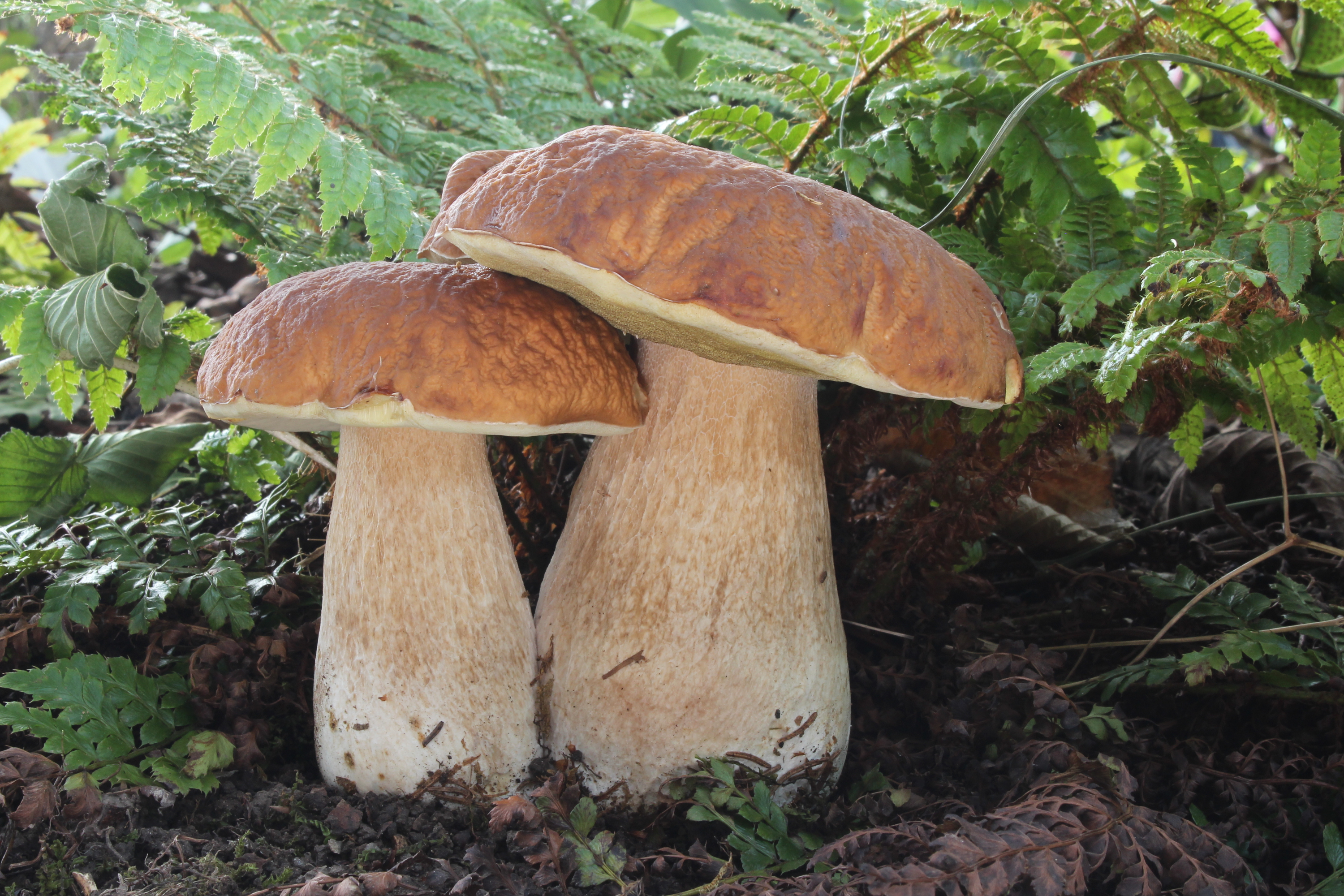
Boletus edulis
Key differences
The bay bolete (Imleria badia) and the king bolete (Boletus edulis) are popular edible mushrooms. The king bolete has a stipe with a light reticulation and pores that are initially white, turning yellow to yellow-olive with age, which do not change color under pressure. Its flesh remains white when sliced. The bay bolete has a brownish stipe without reticulation, and the pores distinctly stain blue under pressure. The flesh of the bay bolete turns blue when sliced, especially in the cap and above the tubes.
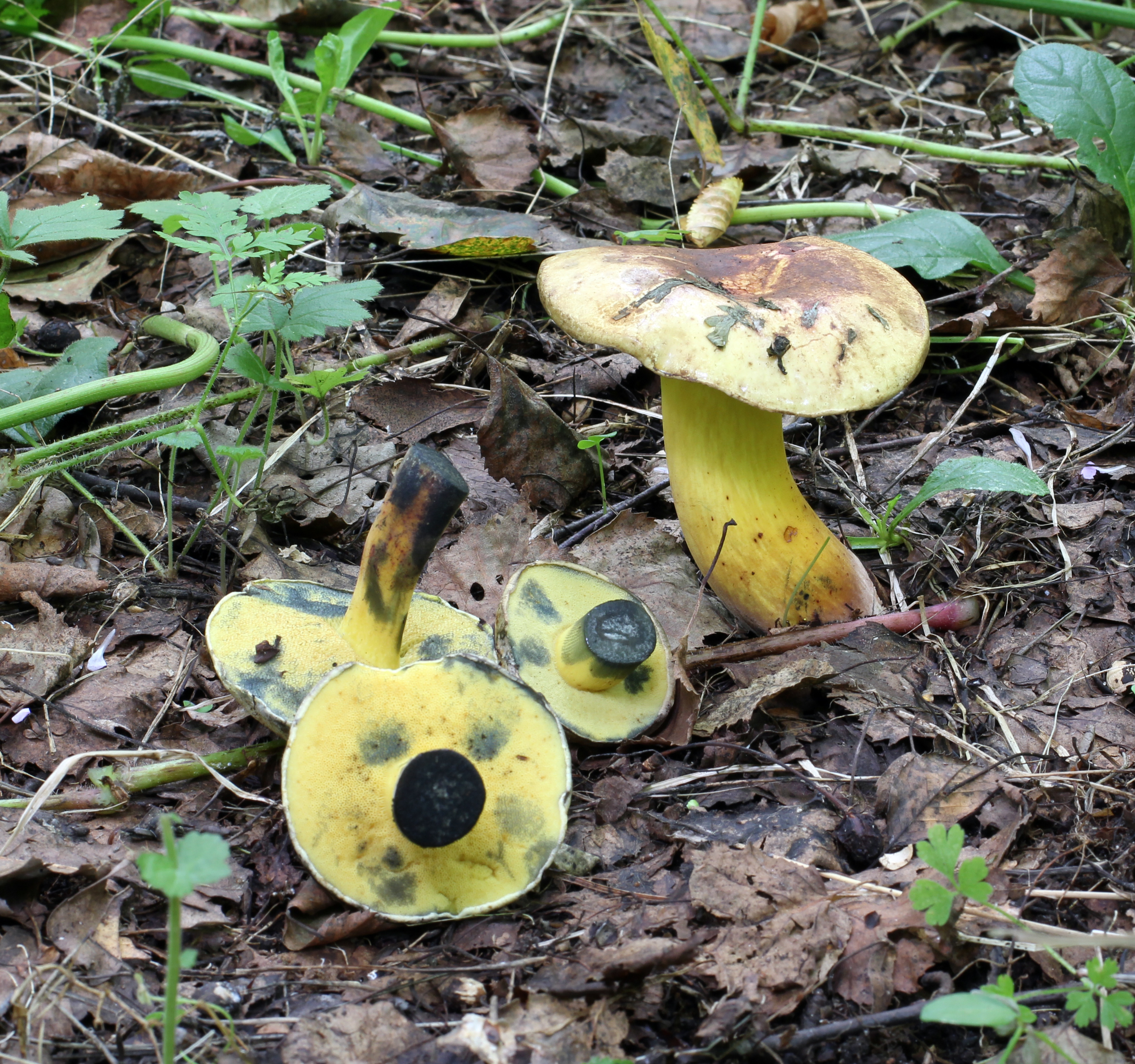
Cyanoboletus pulverulentus
Key differences
The inkstain bolete (Cyanoboletus pulverulentus) reacts to touch and damage with an immediate, rapid, and intense discolouration of all parts of the fruit body to a deep navy-black colour. In Imleria badia, the bluing is less uniform and is concentrated primarily above the tubes and in the upper part of the stipe.
Tylopilus felleus
Key differences
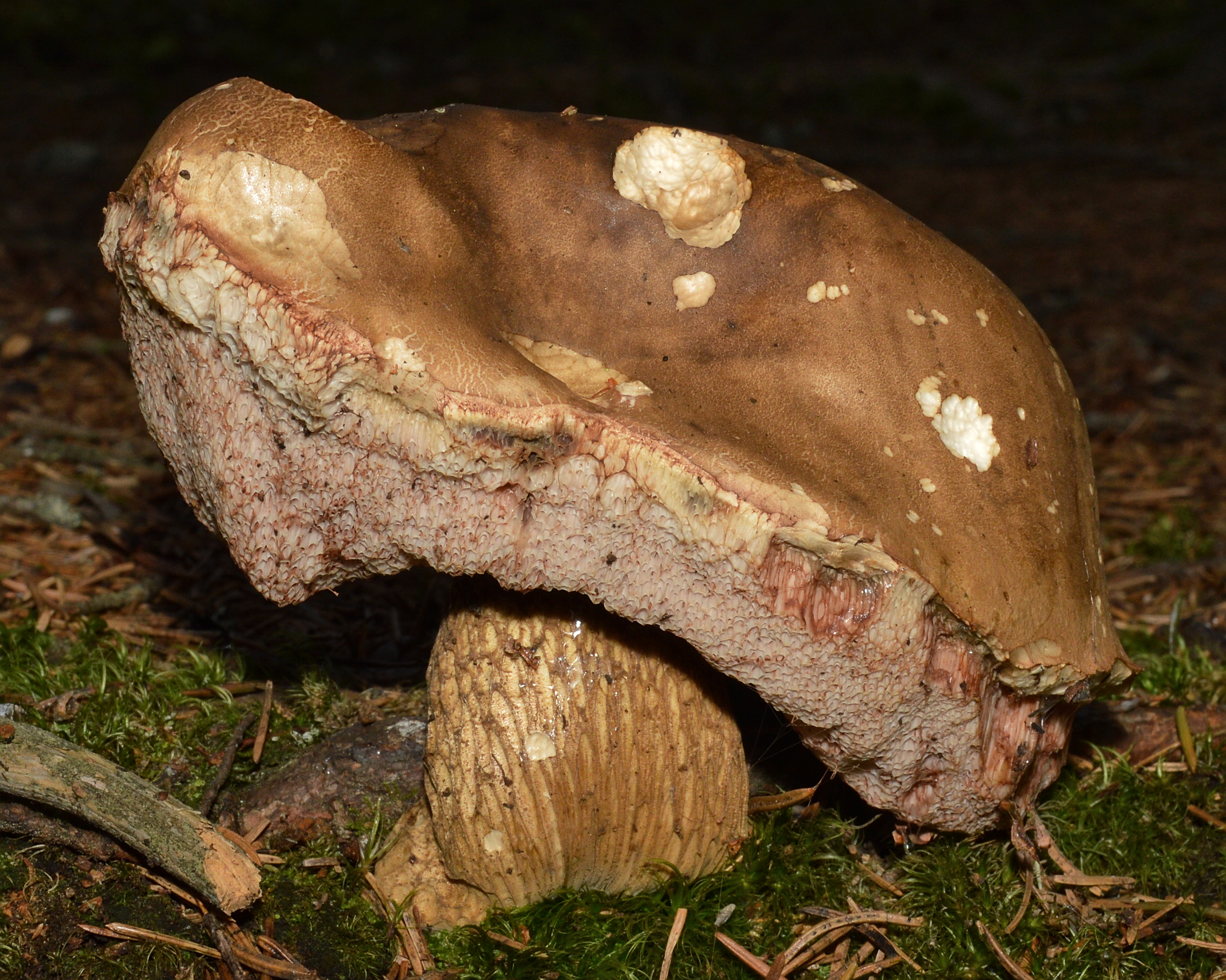
The bitter bolete (Tylopilus felleus) has pores and tubes that are initially white and turn pinkish with age, a distinct dark reticulation on the stipe, and strongly bitter-tasting flesh, making it an inedible mushroom.
The bay bolete (Imleria badia) is sometimes confused with young, darker fruit bodies of the bitter bolete. The bay bolete, however, has yellowish tubes (in the bitter bolete they are white to pinkish), and the stipe lacks the coarse, dark mesh with raised ridges characteristic of the bitter bolete.
Xerocomellus pruinatus
Key differences
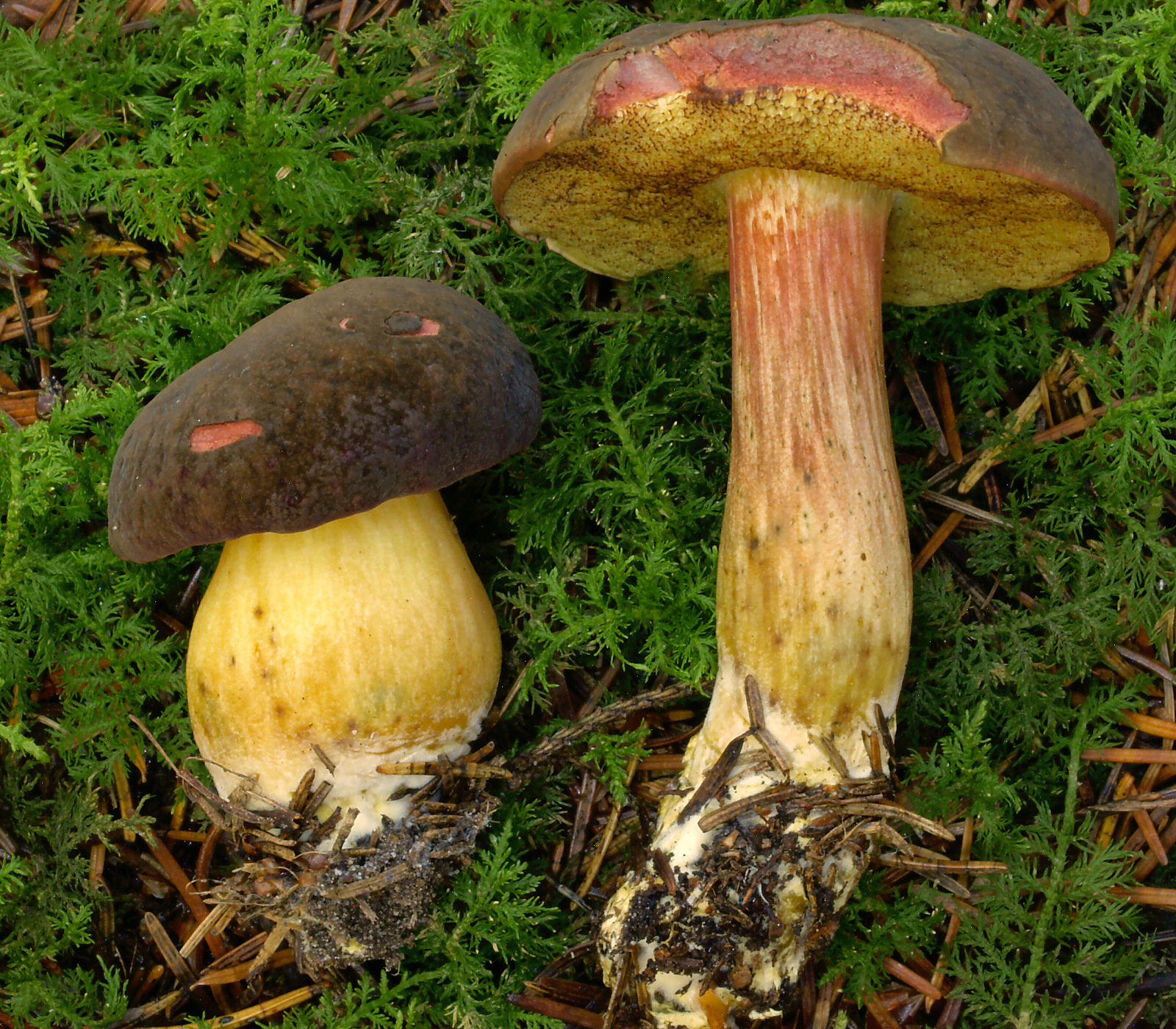
The matt bolete (Xerocomellus pruinatus) can resemble the bay bolete in cap coloration.
FAQ
How to distinguish the bay bolete from the bitter bolete?
The bitter bolete has a prominent dark reticulation on the stem and tubes that turn pink with age and do not bruise blue. The bay bolete lacks a reticulation on the stem, and its tubes are yellowish and immediately bruise blue when pressed.
Does the bay bolete bruise blue?
Yes. Upon pressure, the tubes immediately turn distinctly blue, and the flesh turns blue above the tubes. This is a natural pigment oxidation reaction; it indicates neither a defect nor poison and disappears after cooking.
Can the bay bolete be cultivated in a garden?
In practice, no. It is a strictly mycorrhizal fungus—it requires an active symbiosis with tree roots (mainly pines). 'Garden mycelium' sold in stores almost never yields real crops in a garden.
Where to look for the bay bolete?
Primarily in pine and spruce forests, less often under oaks or beeches. It favors acidic, sandy soils, carpets of green moss, and the edges of heathlands, forming mycorrhizae with trees.
Are podgrzyb brunatny and podgrzybek brunatny the same species?
Yes, it is the same mushroom (Imleria badia). 'Podgrzyb' is the official mycological term, while 'podgrzybek' is the traditional name used by mushroom pickers. In old field guides, it appeared as Boletus badius and Xerocomus badius.
When does the bay bolete grow in Poland?
The peak flush occurs in autumn: from September to the end of October, especially after plentiful rains and cooler nights. The first specimens appear as early as June, and the season closes with frosts.
Does a slippery cap on a bay bolete indicate spoilage?
No. In rain, the skin of the bay bolete naturally becomes sticky and slimy, while in dry weather it is matte. Spoilage is indicated only by spongy, soft flesh, a white coating of mold, or a musty odor.
Enthusiast
Underside of the cap
- Pore diameter
- Pore diameter: 0.5–1.5 mm.
- Tube length
- Tube length: up to 20 mm.
- Pore structure
- Pore structure: compound (tubes).
Names and history
- Obowiązująca nazwa krajowa
- podgrzyb brunatny
- Obowiązująca nazwa taksonomiczna
- Imleria badia (Fr.) Vizzini
- Inne nazwy krajowe
- czarny łebek, czarny łepek, podgrzyb, podsosnówka
- Dawne nazwy krajowe
- podgrzybek brunatny
- Pozostałe nazwy
- Bay Bolete
Home observations
Spore print
- Required materials
- fresh, mature cap, sheet of paper, glass or small bowl, water
- Time
- 2–24 h
- Procedure
- Place the cap underside down on the paper.
- Add a drop of water on top and cover.
- After 2–24 hours, lift the cap and examine the deposit.
What to look for
- Spore print colour
- Spore print colour: brown.
- Spore print shade
- Spore print shade: olive-brown.
Under the microscope
- Required materials
- light microscope, microscope slide and cover slip, needle, water, spores from print
- Procedure
- Transfer a small amount of the spore print onto the slide using a needle.
- Add a drop of water, cover with a cover slip, and examine the spores.
What to look for
- Spore dimensions
- Length 11–18 µm, width 4–6 µm.
Compare dimensions after calibrating the microscope scale.
Reagent tests
- Required materials
- light microscope, microscope slide and cover slip, needle, spores from print, ready-made 5% KOH solution, blotting paper, safety goggles and gloves for working with KOH
- Procedure
- Transfer a small amount of spores from the spore print onto a clean microscope slide.
- Add a drop of ready-made 5% KOH solution and cover with a coverslip.
- Remove excess liquid at the edge of the slide with blotting paper. Observe the spore color under a microscope.
What to look for
- Spore color in 5% KOH
- pale olive-brown
Ecology and distribution
- Soil reaction
- Soil reaction: acidic.
- Soil texture
- Soil texture: sandy.
- Broadleaf trees in the vicinity
- Broadleaf trees in the vicinity: oak.
Broadleaf trees in the vicinity: alder.
Broadleaf trees in the vicinity: sweet chestnut.
Broadleaf trees in the vicinity: hornbeam.
Broadleaf trees in the vicinity: beech. - Coniferous trees in the vicinity
- Coniferous trees in the vicinity: spruce.
Coniferous trees in the vicinity: fir.
Coniferous trees in the vicinity: pine.
Coniferous trees in the vicinity: hemlock.
Coniferous trees in the vicinity: Douglas firs.
Coniferous trees in the vicinity: thujas. - Nearby ground-layer plants
- Nearby ground-layer plants: mosses.
- Mycorrhizal hosts
- greenhouse experiment, Scotland, Isle of Bute, United Kingdom, 2024, field fruiting unconfirmed: Mycorrhizal hosts: Pinus sylvestris (controlled experiment).
General species association: Mycorrhizal hosts: Pinus (general partner list).
General species association: Mycorrhizal hosts: Picea (general partner list).
General species association: Mycorrhizal hosts: Tsuga (general partner list).
General species association: Mycorrhizal hosts: Fagus (general partner list).
General species association: Mycorrhizal hosts: Quercus (general partner list).
General species association: Mycorrhizal hosts: Castanea (general partner list).
General species association: Mycorrhizal hosts: Abies alba (general partner list).
General species association: Mycorrhizal hosts: Pseudotsuga menziesii (general partner list).
General species association: Mycorrhizal hosts: Carpinus betulus (general partner list).
History and culture
The bay bolete occupies an important place in the mushroom-picking traditions of Central Europe and in the history of mycological taxonomy. In Polish folk and regional nomenclature, numerous traditional terms referred to the appearance of the fruit bodies, such as "czarny łepek" (black cap), "kasztaniak" (chestnut bolete), "grzyb gniady" (bay mushroom)—referring to the dark brown colour of the cap—or "siniak" (bruiser), related to the phenomenon of blueing of bruised tubes and flesh.
In the history of scientific mycology, the species underwent a series of significant classification changes reflecting the evolution of bolete systematics. It was originally treated in 1818 by Elias Magnus Fries as Boletus castaneus β badius. Subsequently, for many decades in the mycological literature, it functioned in the genus Xerocomus as Xerocomus badius, into which Édouard-Jean Gilbert transferred it in 1931. In 2014, Italian mycologist Alfredo Vizzini, drawing on modern molecular phylogenetic research, erected the new genus Imleria (named in honour of the Belgian researcher Louis Imler) and transferred the taxon under the name Imleria badia, thereby confirming its distinct evolutionary position within the family Boletaceae.
Edibility and safety
The bay bolete is a valued edible mushroom with a delicate, pleasant aroma and a mild flavour with a discernible forest undertone. An absolute prerequisite for the safe consumption of this species is subjecting the fruit bodies to thermal processing—boiling, frying, or stewing. It must be emphasized that drying the fruit bodies serves merely as a preservation method for the raw material and does not substitute for heat treatment; dried mushrooms must also be subjected to boiling or other appropriate thermal processing prior to consumption. Consuming raw or undercooked fruit bodies poses a risk of digestive disorders.
A specific and significant health safety issue associated with this species is its high capacity for bioaccumulating radioactive elements and metals. Studies published in 2026 (Roczniki Państwowego Zakładu Higieny) documented a pronounced accumulation of radiocaesium (caesium-137) in bay bolete fruit bodies originating from contaminated forest areas. Consequently, special caution and avoiding the foraging of mushrooms in areas subject to historical radioactive contamination are recommended. Old and overripe fruit bodies are likewise unsuitable for consumption.
Key substances and their effects
Norbadione A and caesium
Podgrzybek brunatny zawiera norbadion A – barwnik wykazujący zdolność do silnego wiązania i kumulowania kationów cezu, w tym promieniotwórczego izotopu ¹³⁷Cs. Stopień akumulacji radionuklidów zależy od lokalnego skażenia podłoża i opadu promieniotwórczego na danym stanowisku.
Habitat and occurrence
The bay bolete (Imleria badia) is characterized by a wide geographic distribution across the Northern Hemisphere. According to a biogeographical and phylogenetic revision (Zhao et al., 2014), this species occurs in Europe, North America, and probably in the Far East of Asia.
This species primarily inhabits coniferous and mixed forests, and is also occasionally encountered in deciduous stands. It forms mycorrhizal associations with various tree species, including Scots pine, Norway spruce, larch, as well as European beech and oaks. It requires acidic substrates that are low in calcium carbonate and well-draining. Fruit bodies emerge on sandy soils, among thick layers of coniferous needle litter, in moss cushions, on heathlands, as well as on heavily decayed wood and rotting stumps of coniferous trees.
According to documented phenological data for Poland and the Czech Republic (tables PL and CZ), the fruiting season of the bay bolete spans the period from June to November (months VI–XI). Regional variation in the occurrence of fruit bodies depends mainly on the presence of coniferous and mixed forests on nutrient-poor, acidic sandy soils.
Habitat, region and season
W Czechach grzyb ten rośnie bardzo licznie od czerwca do listopada, niekiedy do początku zimy, przede wszystkim pod świerkami, a w drzewostanach liściastych głównie pod dębami. Owocniki nie wykazują odporności na mróz.
Gatunek jest szeroko rozprzestrzeniony w Europie i Ameryce Północnej, od nizin po regiel górny i granicę lasu. Jako partnerów mikoryzowych wymienia się drzewa iglaste z rodzajów Abies, Picea, Pinus, Pseudotsuga, Thuja i Tsuga, a także liściaste z rodzajów Alnus, Castanea, Carpinus, Fagus i Quercus. Dane molekularne ITS wskazują również na prawdopodobne występowanie w Azji Wschodniej i Afryce Południowej.
Researcher
Edibility and culinary use
Ustalenia źródłowe: jadalność, spożycie i wykorzystanie
- Edibility
- Edible — źródło oznaczenia
Źródło bez opisu bibliograficznego · Retained UTF-8 HTML, line 554; jadalne czy trujące?
- Culinary rating
- Very tasty
Źródło bez opisu bibliograficznego · Retained windows-1250 HTML, line 129; Kuchyňské využití
- Commercial trade
- Approved for trade
Obszar: PL
Zał.1 lp.50; pozostałe wymagania aktu, w tym atest, zachowane.
W Polsce jest dopuszczony do obrotu handlowego oraz do produkcji przetworów grzybowych.
Tekst jednolity rozporządzenia Ministra Zdrowia; Dz.U.2026 poz.258 · PDF str.2–3: § 2–6; str.5–7: zał.1 lp.1–72; status ELI obowiązujący
Scientific names and taxonomy
- Current scientific name
Imleria badia (Fr.) Vizzini
- Former scientific names
- 1818 — Boletus castaneus β badius Fr.
1821 — Boletus badius (Fr.) Fr.
1828 — Boletus castaneus var. badius (Fr.) Fr.
1836 — Boletus glutinosus Krombh.
1838 [1836–1838] — Boletus vaccinus Fr.
1874 [1874–1875] — Boletus limatulus Frost
1881 — Rostkovites badius (Fr.) P. Karst.
1882 — Tubiporus vaccinus (Fr.) P. Karst.
1882 — Tubiporus vaccinus var. ebulbis Fr. ex P. Karst.
1886 — Dictyopus aereus var. vaccinus (Fr.) Quél.
1886 — Viscipelles badia (Fr.) Quél.
1888 — Ixocomus badius (Fr.) Quél.
1898 — Suillus badius (Fr.) Kuntze
1898 — Suillus limatulus (Frost) Kuntze
1898 — Suillus vaccinus (Fr.) Kuntze
1912 [1911] — Boletus badius var. glutinosus (Krombh.) Smotl.
1931 — Xerocomus badius (Fr.) E.-J. Gilbert
1945 — Xerocomus badius var. limatulus (Frost) Snell
1960 — Xerocomus badius f. labyrinthicus Wichanský
1976 — Boletus badius var. glaber Grund & K.A. Harrison
1976 — Boletus badius var. macrostipitatus Grund & K.A. Harrison
2007 — Xerocomus badius f. vaccinus (Fr.) Klofac
2016 — Imleria badia f. vaccina (Fr.) Klofac
2018 — Imleria badia var. limatula (Frost) Blanco-Dios[23]
Obecnie przyjęta nazwa naukowa to Imleria badia (Fr.) Vizzini. Gatunek należy do rodziny Boletaceae, rzędu Boletales oraz klasy Agaricomycetes. Bazonimem jest Boletus castaneus β badius Fr. z 1818 roku. Alfredo Vizzini utworzył rodzaj Imleria i opublikował nową kombinację 12 maja 2014 roku w Index Fungorum 147; taksonomiczne numery rejestracyjne to odpowiednio 550568 i 550569.
[23] [30] [32]W dawniejszym piśmiennictwie podgrzybek brunatny opisywany był głównie jako Boletus badius oraz Xerocomus badius. Dla nazwy Boletus badius rejestr Species Fungorum podaje rok 1821, rewizja Zhu i współautorów wskazuje 1828, a atlas czeski – 1832, przy czym źródła te wymieniają odmienne publikacje pierwotnego opisu. W przypadku nazwy Imleria badia wszystkie trzy opracowania zgodnie podają autorstwo Vizziniego z 2014 roku. Z kolei przy Boletus castaneus var. badius Species Fungorum podaje rok 1828, natomiast rewizja wskazuje 1821.
[23] [32] [13]Rewizja przeprowadzona przez Zhu i współautorów w 2014 roku wyodrębniła rodzaj Imleria z wąsko ujmowanych rodzajów Xerocomus, Xerocomellus i Boletus. W obrębie Imleria wyróżniono cztery gatunki: Imleria badia, Imleria obscurebrunnea, Imleria parva oraz Imleria subalpina. Dawniejsze chińskie doniesienia pod nazwą Boletus badius odnosiły się do trzech pozostałych gatunków, stąd ich dane morfologiczne i ekologiczne nie dotyczą bezpośrednio podgrzybka brunatnego.
[30] [32]W rewizji filogenetycznej analizowano pięć markerów DNA: ITS, nrLSU, tef1α, rpb1 oraz rpb2. Wielogenowa analiza obejmowała regiony 5.8S, nrLSU, tef1α, rpb1 i rpb2, a region ITS badano również osobno. Łącznie dla badanych taksonów uzyskano 55 nowych sekwencji: 13 ITS, 13 nrLSU, 11 tef1α, 10 rpb1 oraz 8 rpb2.
[32]Wielogenowy zbiór danych obejmował 45 taksonów i 4802 pozycje, natomiast oddzielna analiza ITS liczyła 26 sekwencji i 645 pozycji. Wsparcie dla kladu Imleria wyniosło BS = 100% i PP = 1,0. Średnia odległość genetyczna ITS pomiędzy europejskimi a północnoamerykańskimi kolekcjami Imleria badia wyniosła 0,0026 i była wyraźnie niższa od najmniejszego międzygatunkowego dystansu w rodzaju, wynoszącego 0,0176, co uzasadniło utrzymanie populacji z obu kontynentów w ramach jednego gatunku.
[32]Epityp gatunku stanowi szwedzki okaz zebrany przez A. Strida 28 sierpnia 1993 roku w Nordingrå (A. Strid 19707, S-F119691). Z epitypu uzyskano sekwencje referencyjne o numerach dostępu: ITS KJ806970, nrLSU KJ806971, tef1α KJ806969, rpb1 KJ806972 oraz rpb2 KJ806973.
[32]Pomiary mikroskopowe w rewizji taksonomicznej prowadzono na suchych okazach rehydratowanych w 5% KOH, a ocenę barw struktur mikroskopowych wykonywano w wodzie i w KOH. Skórkę kapelusza badano na promienistych przekrojach pionowych w połowie promienia kapelusza, a łuseczki trzonu pobierano z połowy jego wysokości. Obserwacje ultrastruktury zarodników wykonano w mikroskopie skaningowym Hitachi S4800 przy napięciu 10,0 kV.
[32]Ecology and distribution
Laboratory diagnostics
Dane ustrukturyzowane
Basidia
Caulocystidia
- Clamp connections
- Clamp connections: absent. [32]
Hymenial cystidia
- Optical appearance of hymenial cystidia
- Optical appearance of hymenial cystidia: hyaline. [32]
- Distribution of hymenial cystidia
- Distribution of hymenial cystidia: scattered. [32]
- Length of jointly described hymenial cystidia
- Length of jointly described hymenial cystidia: 35–65 µm. [32]
- Shape of jointly described hymenial cystidia
- Shape of jointly described hymenial cystidia: lanceolate, nearly spindle-shaped with a short point or ventricose. [32]
- Wall thickness of jointly described hymenial cystidia
- Wall thickness of jointly described hymenial cystidia: thin-walled. [32]
- Width of jointly described hymenial cystidia
- Width of jointly described hymenial cystidia: 6–10 µm. [32]
Mycorrhiza
- Mycorrhizal branching
- Mycorrhizal branching: dichotomous or unbranched. [29]
- Mycorrhiza length
- Mycorrhiza length: 2–10 mm. [29]
- Abundance of emanating mycorrhizal hyphae
- Abundance of emanating mycorrhizal hyphae: sparse. [29]
- Pattern of the Hartig net
- Pattern of the Hartig net: Palmetti type. [29]
- Appearance of the mycorrhizal mantle
- Appearance of the mycorrhizal mantle: silvery. [29]
- Structure of the mycorrhizal mantle
- Structure of the mycorrhizal mantle: plectenchymatous. [29]
- Surface relief of the mycorrhizal mantle
- Surface relief of the mycorrhizal mantle: smooth. [29]
- Colour of the mycorrhizal rhizomorphs
- Colour of the mycorrhizal rhizomorphs: whitish. [29]
- Presence of mycorrhizal rhizomorphs
- Presence of mycorrhizal rhizomorphs: present. [29]
Pileipellis
- Colour of pileipellis hyphae
- Colour of pileipellis hyphae: yellowish. [32]
- Incrustation of cuticle hyphae
- Incrustation of cuticle hyphae: present. [32]
- Embedding of cuticle hyphae in a gelatinous substance
- Embedding of cuticle hyphae in a gelatinous substance: present. [32]
- Color of cuticle pigment
- Color of cuticle pigment: yellow-brown. [32]
- Structure of the pileipellis
- in young fruit bodies: Structure of the pileipellis: ixotrichoderm. [32]
in mature fruit bodies: Structure of the pileipellis: interwoven ixotrichoderm. [32] - Shape of pileipellis terminal cell apex
- Shape of pileipellis terminal cell apex: rounded. [32]
Shape of pileipellis terminal cell apex: tapering. [32] - Pileipellis terminal cell length
- Pileipellis terminal cell length: 30–60 µm. [32]
- Shape of the terminal cell of the pileipellis
- Shape of the terminal cell of the pileipellis: cylindrical. [32]
- Pileipellis terminal cell width
- Pileipellis terminal cell width: 4–7 µm. [32]
Reactions with reagents
DNA sequences
- Sequence
- Imleria badia; ITS; KJ806970; S-F119691(epitype)
- Sequence
- Imleria badia; nrLSU; KJ806971; S-F119691(epitype)
- Sequence
- Imleria badia; tef1alpha; KJ806969; S-F119691(epitype)
- Sequence
- Imleria badia; rpb1; KJ806972; S-F119691(epitype)
- Sequence
- Imleria badia; rpb2; KJ806973; S-F119691(epitype)
- Sequence
- Imleria badia; rDNA58S; KC215204; HKAS53502
- Sequence
- Imleria badia; nrLSU; KC215213; HKAS53502
- Sequence
- Imleria badia; tef1alpha; KC215247; HKAS53502
- Sequence
- Imleria badia; rpb1; KC215226; HKAS53502
- Sequence
- Imleria badia; rpb2; KC215235; HKAS53502
- Sequence
- Imleria badia; nrLSU; KC215212; HKAS74714
- Sequence
- Imleria badia; tef1alpha; KC215242; HKAS74714
- Sequence
- Imleria badia; rpb1; KC215224; HKAS74714
Spores
- Spore colour under the microscope
- Spore colour under the microscope: nearly hyaline. [32]
- Droplets inside the spore
- Droplets inside the spore: 1–2. [32]
- Spore length-to-width ratio
- Spore length-to-width ratio: 2.3–4.2. [32]
- Spore shape
- Spore shape: nearly spindle-shaped. [32]
Spore shape: ellipsoid. [32]
Spore shape: elongate ellipsoid. [32] - Spore surface
- Spore surface: smooth. [32]
- Spore wall thickness
- Spore wall thickness: 0.5 µm. [32]
Hymenophoral trama
Material and research methods
Key substances and their effects
Próbki marynat odsączono, zamrożono, poddano liofilizacji i rozdrobnieniu, a bezpośrednio przed pomiarem suszono sublimacyjnie przez trzy dni. Aktywność gamma mierzono spektrometrem z detektorem germanowym HPGe o wydajności względnej 18% i rozdzielczości 1,9 keV przy energii 1,332 MeV. Wyniki przeliczono na moment pobrania próbek, a zawartość potasu obliczono na podstawie aktywności ⁴⁰K.
[22]- Results as recorded in the sources — Spore length
- Materiał badania: Wynik atlasowy wymiarów zarodników Imleria badia; badany materiał i konkretna kolekcja nie są podane. 12–16 µm; zakres pomiaru: korpus [13]
Materiał badania: Materiał europejski Imleria badia z opracowania Zhu i in. 2014; wynik opisowy na poziomie badanego zakresu, bez przypisania do pojedynczego vouchera. Materiał suszony, ponownie nawodniony (przygotowanie opisano w Metodach): Zarodnik może mieć długość od 11 do 17,5 mikrometra. [32]
Materiał badania: Wynik atlasowy wymiarów zarodników Imleria badia; badany materiał i konkretna kolekcja nie są podane. 11–18 µm; zakres pomiaru: korpus [24] - Results as recorded in the sources — Spore width
- Materiał badania: Materiał europejski Imleria badia z opracowania Zhu i in. 2014; wynik opisowy na poziomie badanego zakresu, bez przypisania do pojedynczego vouchera. Materiał suszony, ponownie nawodniony (przygotowanie opisano w Metodach): Zarodnik może mieć szerokość od 3,5 do 5 mikrometrów. [32]
Materiał badania: Wynik atlasowy wymiarów zarodników Imleria badia; badany materiał i konkretna kolekcja nie są podane. 4,5–6 µm; zakres pomiaru: korpus [24]
Materiał badania: Wynik atlasowy wymiarów zarodników Imleria badia; badany materiał i konkretna kolekcja nie są podane. 4–5,5 µm; zakres pomiaru: korpus [13]
Metody i zapisy źródłowe — Materiał europejski Imleria badia z opracowania Zhu i in. 2014; wynik opisowy na poziomie badanego zakresu, bez przypisania do pojedynczego vouchera.
Stan materiału: ponownie nawodniony.
Pochodzenie materiału: Badane europejskie kolekcje rewizji SE/DE/IT; nie wynik z Polski..
Identyfikacja kropli: nierozstrzygnięta.
Metoda źródłowa. Technika: mikroskopia świetlna (LM). Medium: KOH. Stężenie: 5%. Opis metody w źródle: Mikroskopia świetlna wysuszonego materiału po rehydratacji w 5% KOH; barwy mikrostruktur badano w wodzie, z zachowaniem jawnego medium KOH przy opisie zarodników. Nie podano kalibracji ani powiększenia. SEM jest osobną opisaną metodą powierzchni bazydiospor, nie metodą wszystkich wyników.
Liczba zarodników na jednej podstawce; Długość podstawki; Szerokość podstawki — zapis źródłowy: „Basidia 25–48 × 8–14 μm, clavate, 4-spored.” [32]
Kształt podstawki — zapis źródłowy: „clavate” [32]
Układ kaulocystyd — zapis źródłowy: „caespitose” [32]
Długość kaulocystyd; Szerokość kaulocystyd — zapis źródłowy: „Caulocystidia 29–65 × 5–9 μm” [32]
Kształt kaulocystyd — zapis źródłowy: „clavate, ventricose” [32]
Wygląd optyczny cystyd hymenialnych — zapis źródłowy: „Pleuro- and cheilocystidia ... hyaline when young” [32]
Rozmieszczenie cystyd hymenialnych — zapis źródłowy: „Pleuro- and cheilocystidia scattered” [32]
Długość wspólnie opisanych cystyd hymenialnych; Szerokość wspólnie opisanych cystyd hymenialnych — zapis źródłowy: „Pleuro- and cheilocystidia scattered, 35–65 × 6–10 μm” [32]
Kształt wspólnie opisanych cystyd hymenialnych — zapis źródłowy: „lanceolate to subfusoid-mucronate or ventricose” [32]
Grubość ściany wspólnie opisanych cystyd hymenialnych — zapis źródłowy: „thin-walled” [32]
Barwa strzępek skórki kapelusza — zapis źródłowy: „yellowish interwoven hyphae” [32]
Inkrustacja strzępek skórki — zapis źródłowy: „slightly incrusted by yellow-brown (4B8) pigment” [32]
Osadzenie strzępek skórki w substancji żelowej — zapis źródłowy: „embedded in gelatinous substance” [32]
Barwa pigmentu skórki — zapis źródłowy: „yellow-brown (4B8) pigments” [32]
Organizacja skórki kapelusza — zapis źródłowy: „ixotrichoderm in youth” [32]
Organizacja skórki kapelusza — zapis źródłowy: „becoming an ixo-intricate-trichoderm when mature” [32]
Kształt wierzchołka komórki końcowej skórki kapelusza; Długość komórki końcowej skórki kapelusza; Kształt komórki końcowej skórki kapelusza; Szerokość komórki końcowej skórki kapelusza — zapis źródłowy: „terminal cells cylindrical with rounded to tapering apex, 30–60 × 4–7μm.” [32]
Barwa zarodnika w mikroskopie — zapis źródłowy: „nearly hyaline to pale olivaceous brown (2B6) in 5% KOH” [32]
Krople wewnątrz zarodnika — zapis źródłowy: „Jedna lub dwie krople w dojrzałych zarodnikach.” [32]
Długość zarodnika — zapis źródłowy: „11–15(17,5) × (3,5)4–5 µm; długość 11–15(17,5) µm; n = 160 / 8 owocników / 5 kolekcji, ≥90% bez nawiasów, rehydratacja 5% KOH.” [32]
Iloraz długości i szerokości zarodnika — zapis źródłowy: „Q = (2,3)2,5–3,6(4,2); Qm = 3,0 ± 0,33 (średnia ± odchylenie standardowe); 160 zarodników / 8 owocników / 5 kolekcji.” [32]
Kształt zarodnika — zapis źródłowy: „Boletoidalne: z boku niemal wrzecionowate, od czoła elipsoidalne do podłużnych.” [32]
Powierzchnia zarodnika — zapis źródłowy: „Gładkie pod mikroskopem świetlnym i skaningowym.” [32]
Grubość ściany zarodnika — zapis źródłowy: „Ściana nieco pogrubiała, około 0,5 µm.” [32]
Szerokość zarodnika — zapis źródłowy: „11–15(17,5) × (3,5)4–5 µm; szerokość (3,5)4–5 µm; n = 160 / 8 owocników / 5 kolekcji, ≥90% bez nawiasów, rehydratacja 5% KOH.” [32]
Metody i zapisy źródłowe — Źródłowy opis gatunku Boletus badius, nie diagnoza całego rodzaju ani własny pomiar kolekcji.
Typ tramy hymenoforu — zapis źródłowy: „Gatunkowa uwaga na s. 33: Boletus badius ma prawdziwie boletoidalną tramę hymenoforu; terminologia na s. 3–4 zalicza ten podtyp do tramy bilateralnej.” [27]
Metody i zapisy źródłowe — Źródłowy opis gatunku Boletus badius, nie diagnoza całego rodzaju ani własny pomiar kolekcji. Lista badanego materiału gatunku na s. 34: JŠ 373, 395, 1814, 1856, 1858, 1875, 1931–32, 1980–81, 2102–03, 3089, 3116, 3120, 3223–25, 4299. Zachowana jako kontekst publikacji, bez wyboru vouchera ani wyliczania N.
Typ tramy hymenoforu — zapis źródłowy: „Na s. 31 B. badius jest wymieniony wprost wśród taksonów z prawdziwie boletoidalną tramą. Wcześniejsza wspólna fraza o B. badius i B. moravicus dopuszcza wygląd boletoidalny lub prawie boletoidalny.” [28]
Metody i zapisy źródłowe — Controlled Imleria badia–Pinus sylvestris mycorrhizal experiment, Bute, Scotland
Pochodzenie materiału: Isle of Bute, Scotland, GB.
Rozgałęzienie mikoryzy — zapis źródłowy: „dichotomous ramification pattern or ramification was absent” [29]
Długość mikoryzy — zapis źródłowy: „2 mm to 10 mm” [29]
Obfitość strzępek wychodzących mikoryzy — zapis źródłowy: „infrequent emanating hyphae” [29]
Wzór sieci Hartiga mikoryzy — zapis źródłowy: „Palmetti-type pattern” [29]
Wygląd płaszczyka mikoryzy — zapis źródłowy: „The mantle was either silvery or smooth” [29]
Organizacja płaszczyka mikoryzy — zapis źródłowy: „plectenchymatous organisation” [29]
Relief powierzchni płaszczyka mikoryzy — zapis źródłowy: „either silvery or smooth” [29]
Barwa ryzomorf mikoryzy — zapis źródłowy: „whitish rhizomorphs” [29]
Obecność ryzomorf mikoryzy — zapis źródłowy: „Rhizomorphs were present” [29]
Barwa nierozgałęzionych zakończeń mikoryzy — zapis źródłowy: „younger unramified ends whitish” [29]
Barwa nierozgałęzionych zakończeń mikoryzy — zapis źródłowy: „older unramified ends ochre, yellowish-brown or whitish” [29]
Metody i zapisy źródłowe — Wynik atlasowy wymiarów zarodników Imleria badia; badany materiał i konkretna kolekcja nie są podane.
Długość zarodnika — zapis źródłowy: „11–18 × 4,5–6 µm; długość 11–18 µm, n i protokół niepodane.” [24]
Szerokość zarodnika — zapis źródłowy: „11–18 × 4,5–6 µm; szerokość 4,5–6 µm, n i protokół niepodane.” [24]
Metody i zapisy źródłowe — Wynik atlasowy wymiarów zarodników Imleria badia; badany materiał i konkretna kolekcja nie są podane.
Długość zarodnika — zapis źródłowy: „12–16 × 4–5,5 µm; długość 12–16 µm, n i protokół niepodane.” [13]
Szerokość zarodnika — zapis źródłowy: „12–16 × 4–5,5 µm; szerokość 4–5,5 µm, n i protokół niepodane.” [13]
Occurrence verification — scope and limitations
Materiał: Pierwotne kolekcje autora atlasu z Dolnego Śląska.
Zakres odczytu: Fragmenty źródeł.
Odczytano fragment źródła; pełny tekst i metody nie zostały zweryfikowane.
Data przeglądu: 21.10.2026.
Ograniczenia: Źródłowe oznaczenie i zakres pojęcia zachowano. Nie zadeklarowano nowego badania okazów ani odczytu wszystkich publikacji cytowanych przez źródło.
[24]- Sample preparation for microscopy
- Materiał suszony, ponownie nawodniony. Mikroskopia świetlna wysuszonego materiału po rehydratacji w 5% KOH; barwy mikrostruktur badano w wodzie, z zachowaniem jawnego medium KOH przy opisie zarodników. Nie podano kalibracji ani powiększenia. SEM jest osobną opisaną metodą powierzchni bazydiospor, nie metodą wszystkich wyników. [32]
Materiał ponownie nawodniony. [32]
Protokół dla opisanych obserwacji: Mikroskopia świetlna wysuszonego materiału po rehydratacji w 5% KOH; barwy mikrostruktur badano w wodzie, z zachowaniem jawnego medium KOH przy opisie zarodników. Nie podano kalibracji ani powiększenia. SEM jest osobną opisaną metodą powierzchni bazydiospor, nie metodą wszystkich wyników. [32]
Scope of the review
Before completion, the record requires completion or settlement.
- Search for occurrence regions requires reconciliation.
- Connections of earlier findings require re-examination.
- New sources require accounting for information encountered during the review.
Legal basis and scope of assessment
Key substances and their effects
W badaniach biochemicznych Gąseckiej i współpracowników (2017) oceniano profil metali, kwasów fenolowych oraz flawonoidów w owocnikach Imleria badia ze stanowisk o różnym stopniu zanieczyszczenia antropogenicznego. Z kolei Sotek i współpracownicy (2025) badali związki bioaktywne, zdolności antyoksydacyjne oraz zawartość pierwiastków śladowych w trzech gatunkach grzybów, w tym u podgrzybka brunatnego.
[18] [26]Analytical results
Imleria badia; drained retail pickled mushrooms; Gdańsk; 2016; dry mass
Imleria badia; drained retail pickled mushrooms; Gdańsk; 2016; wet mass
Boletus edulis; pooled experimental material; caps; fresh → dried; near Osiek in the Tuchola Pinewoods, north-central Poland; 2015; dry mass
Boletus edulis; pooled experimental material; caps; fresh; near Osiek in the Tuchola Pinewoods, north-central Poland; 2015; wet mass
Boletus edulis; pooled experimental material; caps; fresh → dried#; near Osiek in the Tuchola Pinewoods, north-central Poland; 2015; dry mass
- total K
- 27000 ± 1100 mg/kg; authors’ calculation; dry-mass basis.[22]
Boletus edulis; pooled experimental material; caps; fresh → dried#; near Osiek in the Tuchola Pinewoods, north-central Poland; 2015; wet mass
- total K
- 2700 ± 110 mg/kg; authors’ calculation; wet-mass basis.[22]
Boletus edulis; pooled experimental material; stipes; fresh → dried; near Osiek in the Tuchola Pinewoods, north-central Poland; 2015; dry mass
Boletus edulis; pooled experimental material; stipes; fresh; near Osiek in the Tuchola Pinewoods, north-central Poland; 2015; wet mass
Boletus edulis; pooled experimental material; stipes; fresh → dried#; near Osiek in the Tuchola Pinewoods, north-central Poland; 2015; dry mass
- total K
- 12000 ± 1800 mg/kg; authors’ calculation; dry-mass basis.[22]
Boletus edulis; pooled experimental material; stipes; fresh → dried#; near Osiek in the Tuchola Pinewoods, north-central Poland; 2015; wet mass
- total K
- 1200 ± 180 mg/kg; authors’ calculation; wet-mass basis.[22]
Boletus edulis; pooled experimental material; whole fruiting bodies; fresh → dried; near Osiek in the Tuchola Pinewoods, north-central Poland; 2015; dry mass
Boletus edulis; pooled experimental material; whole mushrooms; fresh; near Osiek in the Tuchola Pinewoods, north-central Poland; 2015; wet mass
Boletus edulis; pooled experimental material; whole mushrooms; fresh → dried#; near Osiek in the Tuchola Pinewoods, north-central Poland; 2015; dry mass
- total K
- 21000 ± 1400 mg/kg; authors’ calculation; dry-mass basis.[22]
Boletus edulis; pooled experimental material; whole mushrooms; fresh → dried#; near Osiek in the Tuchola Pinewoods, north-central Poland; 2015; wet mass
- total K
- 2100 ± 140 mg/kg; authors’ calculation; wet-mass basis.[22]
Boletus edulis; pooled experimental material; caps; fresh → blanched; near Osiek in the Tuchola Pinewoods, north-central Poland; 2015; dry mass
Boletus edulis; pooled experimental material; caps; fresh → blanched; near Osiek in the Tuchola Pinewoods, north-central Poland; 2015; wet mass
Boletus edulis; pooled experimental material; stipes; fresh → blanched; near Osiek in the Tuchola Pinewoods, north-central Poland; 2015; dry mass
Boletus edulis; pooled experimental material; stipes; fresh → blanched; near Osiek in the Tuchola Pinewoods, north-central Poland; 2015; wet mass
Boletus edulis; pooled experimental material; whole fruiting bodies; fresh → blanched; near Osiek in the Tuchola Pinewoods, north-central Poland; 2015; dry mass
Boletus edulis; pooled experimental material; whole mushrooms; fresh → blanched; near Osiek in the Tuchola Pinewoods, north-central Poland; 2015; wet mass
Boletus edulis; pooled experimental material; whole mushrooms; fresh → blanched; near Osiek in the Tuchola Pinewoods, north-central Poland; 2015; dry mass
- total K
- 14000 ± 2200 mg/kg; authors’ calculation; dry-mass basis.[22]
Boletus edulis; pooled experimental material; caps; fresh → blanched → pickled; near Osiek in the Tuchola Pinewoods, north-central Poland; 2015; dry mass
Boletus edulis; pooled experimental material; caps; fresh → blanched → pickled; near Osiek in the Tuchola Pinewoods, north-central Poland; 2015; wet mass
Boletus edulis; pooled experimental material; stipes; fresh → blanched → pickled; near Osiek in the Tuchola Pinewoods, north-central Poland; 2015; dry mass
Boletus edulis; pooled experimental material; stipes; fresh → blanched → pickled; near Osiek in the Tuchola Pinewoods, north-central Poland; 2015; wet mass
Boletus edulis; pooled experimental material; whole fruiting bodies; fresh → blanched → pickled; near Osiek in the Tuchola Pinewoods, north-central Poland; 2015; dry mass
Boletus edulis; pooled experimental material; whole mushrooms; fresh → blanched → pickled; near Osiek in the Tuchola Pinewoods, north-central Poland; 2015; wet mass
Boletus edulis; pooled experimental material; whole mushrooms; fresh → blanched → pickled; near Osiek in the Tuchola Pinewoods, north-central Poland; 2015; dry mass
- total K
- 2700 ± 1000 mg/kg; authors’ calculation; dry-mass basis.[22]
Boletus edulis; pooled experimental material; caps; fresh → dried (powdered) → macerated; near Osiek in the Tuchola Pinewoods, north-central Poland; 2015; dry mass
Boletus edulis; pooled experimental material; caps; fresh → dried (powdered) → macerated; near Osiek in the Tuchola Pinewoods, north-central Poland; 2015; wet mass
Boletus edulis; pooled experimental material; caps; fresh → dried (ground) → macerated; near Osiek in the Tuchola Pinewoods, north-central Poland; 2015; dry mass
- total K
- 3200 ± 1800 mg/kg; authors’ calculation; dry-mass basis.[22]
Boletus edulis; pooled experimental material; caps; fresh → dried (ground) → macerated; near Osiek in the Tuchola Pinewoods, north-central Poland; 2015; wet mass
- total K
- 320 ± 180 mg/kg; authors’ calculation; wet-mass basis.[22]
Boletus edulis; pooled experimental material; stipes; fresh → dried (powdered) → macerated; near Osiek in the Tuchola Pinewoods, north-central Poland; 2015; dry mass
Boletus edulis; pooled experimental material; stipes; fresh → dried (powdered) → macerated; near Osiek in the Tuchola Pinewoods, north-central Poland; 2015; wet mass
Boletus edulis; pooled experimental material; stipes; fresh → dried (ground) → macerated; near Osiek in the Tuchola Pinewoods, north-central Poland; 2015; dry mass
- total K
- 1500 ± 800 mg/kg; authors’ calculation; dry-mass basis.[22]
Boletus edulis; pooled experimental material; stipes; fresh → dried (ground) → macerated; near Osiek in the Tuchola Pinewoods, north-central Poland; 2015; wet mass
- total K
- 150 ± 80 mg/kg; authors’ calculation; wet-mass basis.[22]
Boletus edulis; pooled experimental material; whole fruiting bodies; fresh → dried (powdered) → macerated; near Osiek in the Tuchola Pinewoods, north-central Poland; 2015; dry mass
Boletus edulis; pooled experimental material; whole mushrooms; fresh → dried (powdered) → macerated; near Osiek in the Tuchola Pinewoods, north-central Poland; 2015; wet mass
Boletus edulis; pooled experimental material; whole mushrooms; fresh → dried (ground) → macerated; near Osiek in the Tuchola Pinewoods, north-central Poland; 2015; dry mass
- total K
- 2500 ± 1400 mg/kg; authors’ calculation; dry-mass basis.[22]
Boletus edulis; pooled experimental material; whole mushrooms; fresh → dried (ground) → macerated; near Osiek in the Tuchola Pinewoods, north-central Poland; 2015; wet mass
- total K
- 250 ± 140 mg/kg; authors’ calculation; wet-mass basis.[22]
Boletus edulis; pooled experimental material; caps; fresh → deep frozen → blanched; near Osiek in the Tuchola Pinewoods, north-central Poland; 2015; dry mass
Boletus edulis; pooled experimental material; caps; fresh → deep frozen → blanched; near Osiek in the Tuchola Pinewoods, north-central Poland; 2015; wet mass
Boletus edulis; pooled experimental material; stipes; fresh → deep frozen → blanched; near Osiek in the Tuchola Pinewoods, north-central Poland; 2015; dry mass
Boletus edulis; pooled experimental material; stipes; fresh → deep frozen → blanched; near Osiek in the Tuchola Pinewoods, north-central Poland; 2015; wet mass
Boletus edulis; pooled experimental material; whole fruiting bodies; fresh → deep frozen → blanched; near Osiek in the Tuchola Pinewoods, north-central Poland; 2015; dry mass
Boletus edulis; pooled experimental material; whole mushrooms; fresh → deep frozen → blanched; near Osiek in the Tuchola Pinewoods, north-central Poland; 2015; wet mass
Boletus edulis; pooled experimental material; whole mushrooms; fresh → deep frozen → blanched; near Osiek in the Tuchola Pinewoods, north-central Poland; 2015; dry mass
- total K
- 8800 ± 1200 mg/kg; authors’ calculation; dry-mass basis.[22]
Boletus edulis; pooled experimental material; caps; fresh → deep frozen → blanched → pickled; near Osiek in the Tuchola Pinewoods, north-central Poland; 2015; dry mass
Boletus edulis; pooled experimental material; caps; fresh → deep frozen → blanched → pickled; near Osiek in the Tuchola Pinewoods, north-central Poland; 2015; wet mass
Boletus edulis; pooled experimental material; stipes; fresh → deep frozen → blanched → pickled; near Osiek in the Tuchola Pinewoods, north-central Poland; 2015; dry mass
Boletus edulis; pooled experimental material; stipes; fresh → deep frozen → blanched → pickled; near Osiek in the Tuchola Pinewoods, north-central Poland; 2015; wet mass
Boletus edulis; pooled experimental material; whole fruiting bodies; fresh → deep frozen → blanched → pickled; near Osiek in the Tuchola Pinewoods, north-central Poland; 2015; dry mass
Boletus edulis; pooled experimental material; whole mushrooms; fresh → deep frozen → blanched → pickled; near Osiek in the Tuchola Pinewoods, north-central Poland; 2015; wet mass
Boletus edulis; pooled experimental material; whole mushrooms; fresh → deep frozen → blanched → pickled; near Osiek in the Tuchola Pinewoods, north-central Poland; 2015; dry mass
- total K
- 3200 ± 850 mg/kg; authors’ calculation; dry-mass basis.[22]
Boletus edulis; drained retail pickled mushrooms; commercially pickled and drained; Gdańsk; 2016; dry mass
Ecological studies
Habitat, region and season
Doświadczenie szklarniowe przeprowadzone na wyspie Bute w Szkocji potwierdziło tworzenie mikoryzy przez Imleria badia na siewkach sosny zwyczajnej (Pinus sylvestris). Nasiona wysiano 20 kwietnia 2024 roku, inokulację przeprowadzono 30 lipca, a ocenę systemu korzeniowego wykonano w listopadzie. Doświadczenie obejmowało 20 siewek w każdym wariancie (do szczegółowej analizy losowano po 10), hodowanych na sterylizowanym podłożu torf–perlit–wermikulit (1:1:1), przy użyciu pojedynczego izolatu oraz trzech metod zakażania: grzybnią, zarodnikami oraz metodą łączoną.
[29]W próbach inokulacji grzybniowej stosowano wycinek agaru o powierzchni 1 cm², natomiast w wariancie zarodnikowym dawkę odpowiadającą około 0,25 g wysuszonego kapelusza na roślinę. Ocenę korzeni prowadzono na trzech poziomach głębokości (0–4 cm, 4–8 cm i 8–12 cm), analizując po trzy losowe fragmenty korzeni z każdej strefy pod mikroskopem stereoskopowym i świetlnym, z barwieniem błękitem bawełnianym w laktofenolu.
[29]Wytworzone ektomikoryzy miały długość 2–10 mm, były nierozgałęzione lub widlasto (dichotomicznie) rozgałęzione. Młode struktury były białawe, starsze ochrowe do żółtobrązowych. Obserwowano białawe ryzomorfy, gładki lub srebrzysty mufkowaty płaszczyk o budowie plektenchymatycznej oraz sieć Hartiga typu Palmetti. W strzępkach i ryzomorfach nie stwierdzono sprzążek. Tożsamość grzyba potwierdzono testem PCR specyficznym dla Imleria badia (produkt o długości 151 par zasad) w 9 niezależnych mikoryzach; korzenie kontrolne dały wynik negatywny.
[29]Średni wskaźnik kolonizacji korzeni wyniósł 7,8% dla grzybni, 4,3% dla zawiesiny zarodników oraz 7,16% dla metody łączonej. Na poszczególnych głębokościach (0–4 cm, 4–8 cm, 8–12 cm) wartości te kształtowały się odpowiednio: 11,7%, 6,3% i 5,4% przy inokulacji grzybnią; 3,3%, 6,4% i 3,2% przy inokulacji zarodnikami; 7,0%, 9,1% i 5,2% przy zastosowaniu obu metod. W grupie kontrolnej nie wykryto mikoryz Imleria badia, a sporadyczna obecność innego grzyba wyniosła średnio 1,11%, 1,33% i 0,89% (łącznie około 1%).
[29]Test Kruskala–Wallisa wykazał statystycznie istotną różnicę pomiędzy grupami inokulowanymi a kontrolą (χ² = 26,77, df = 3, p = 6,57 × 10⁻⁶). Testy Wilcoxona z poprawką Bonferroniego potwierdziły istotną kolonizację względem kontroli dla inokulum grzybniowego (p = 4 × 10⁻⁶), metody łączonej (p = 0,00077) oraz zarodników (p = 0,00031). Pomiędzy samymi metodami inokulacji nie stwierdzono istotnych różnic statystycznych (p > 0,05). Podobnie głębokość podłoża nie wpływała istotnie na poziom kolonizacji (p = 0,3497, 0,3668 i 0,9994).
[29]Czysta kultura jednego z izolatów tworzyła zaczątki owocników (primordia) na pożywkach MMN i PDA po przeniesieniu z inkubatora o temperaturze 23 °C do warunków laboratoryjnych o temperaturze 14–18 °C na okres kilku tygodni. Badania te dotyczą wczesnych etapów formowania mikoryzy i zawiązków, nie stanowiąc metody komercyjnej uprawy owocników.
[29]Analiza biogeograficzna oparta na bazie danych GBIF objęła 69 737 globalnych rekordów występowania Imleria badia i jej synonimów, z których wyselekcjonowano 34 151 rekordów europejskich zgromadzonych w latach 1970–2024 (1970–2025). Dane te reprezentują zgłoszenia obecności gatunku na stanowiskach.
[29]Habitat and occurrence
Potwierdzony zasięg występowania
Wszystkie obszary potwierdzone w zapisanych danych źródłowych.
| Kraj | Regiony i stanowiska | Owocnikowanie | Sources |
|---|---|---|---|
| Czechia | Nie ustalono regionów w zapisanych danych | VI–XI |
|
| Poland | Lower Silesia (area named in the source) | VI–XI |
|
Materials
Study documentation
- Microscopy
- src-cms-badia[13]
- Taste description
- src-coford 2008-badia[14]
- Quantitative analysis
- Saba M., Falandysz J. (2021), Environ. Sci. Pollut. Res. 28:12441–12446; DOI 10.1007/s11356-020-11147-7[22]
- Microscopy
- src-snowarski-badia[24]
- Microscopy
- src-sutara 2005[27]
- Microscopy
- src-thomas 2026[29]
- Reagent tests
- Reagent tests
- Reagent tests
- Reagent tests
- Clamp connection study
- src-zhu 2014[32]
- Microscopy
- src-zhu 2014[32]
- Phylogenetic study
- src-zhu 2014[32]
Herbarium collections
- S-F119691(epitype)
Sweden; Angermanland,Nordingra; 1993-08-28; A.Strid19707
[32]- HKAS74714
Germany; Marburg; 2006-12-04; Z.L.YangM3
[32]- HKAS53502
Germany; Marburg; 2006-11-24; L.Wang4
[32]- HKAS74713
Italy; Lazio,CaldaradiManziana/Rome; 2010-11-25; M.GelardiMG396a; 265 m above sea level
[32]- HKAS75797
Italy; Lombardy,Cevo/Brescia; 2012-08-20; M.GelardiMG463a; 1750 m above sea level
[32]
Sources
- Weryfikacja strukturalnych polskich wyników prawnych, 17.10.2026
- src-cms-badia
- src-coford 2008-badia
- ELI: protection
- ELI: trade
- src-gasecka 2017-access
- Saba M., Falandysz J. (2021), Environ. Sci. Pollut. Res. 28:12441–12446; DOI 10.1007/s11356-020-11147-7
- src-sf-badia
- src-snowarski-badia
- src-sotek 2025-access
- src-sutara 2005
- src-sutara 2008
- src-thomas 2026
- src-vizzini 2014
- src-zhu 2014