Edible
Requires preparation

Set your location to see data for your region.
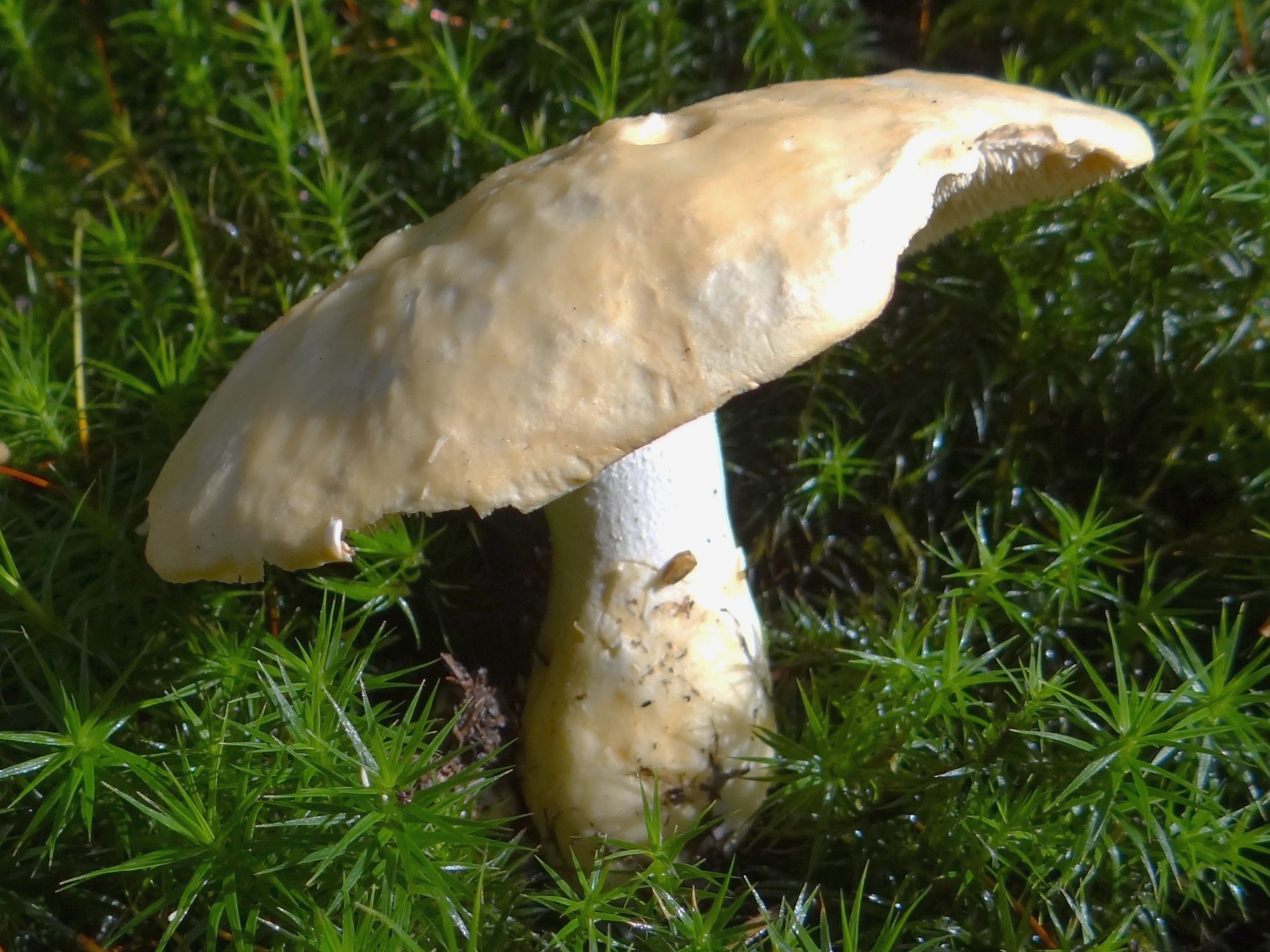

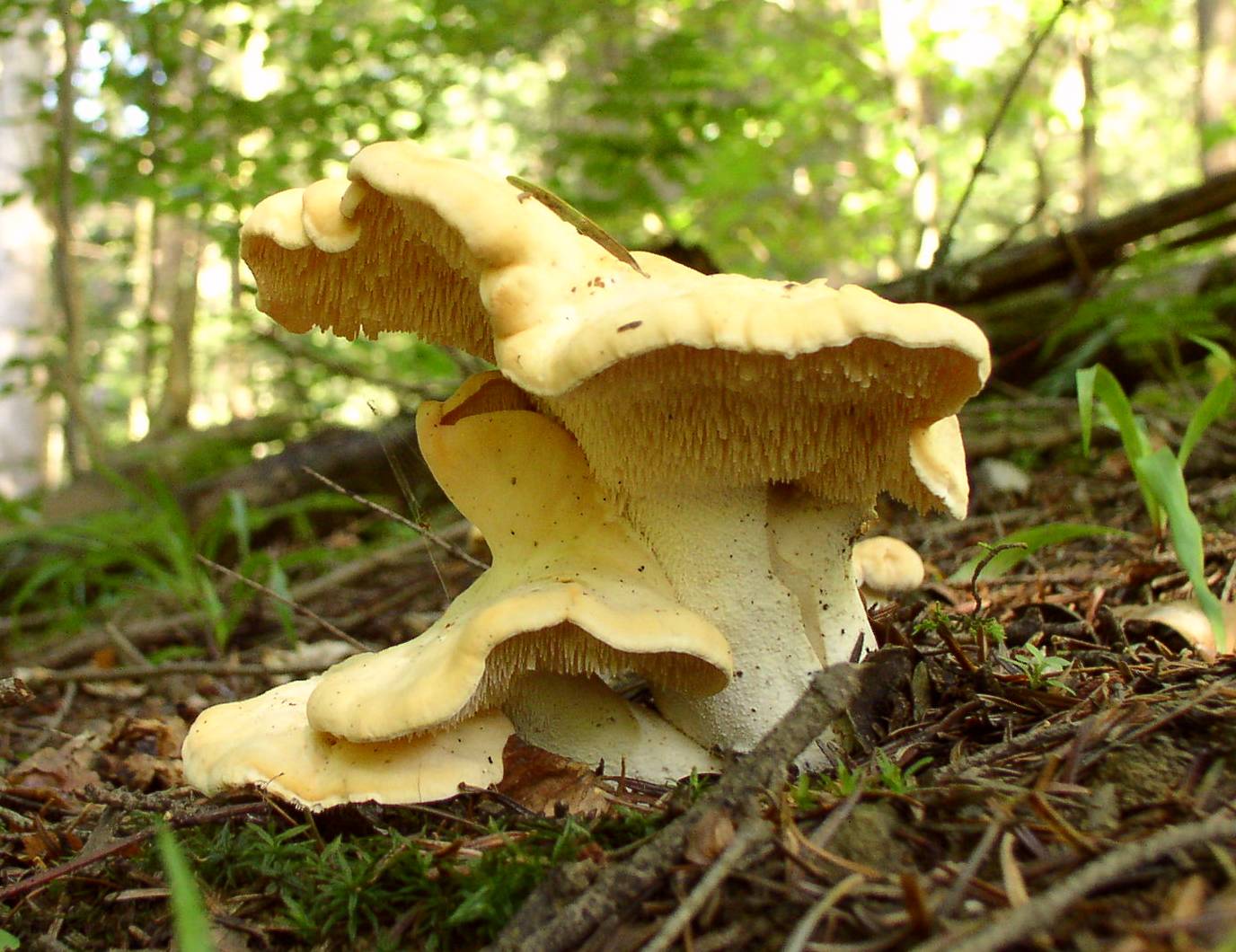

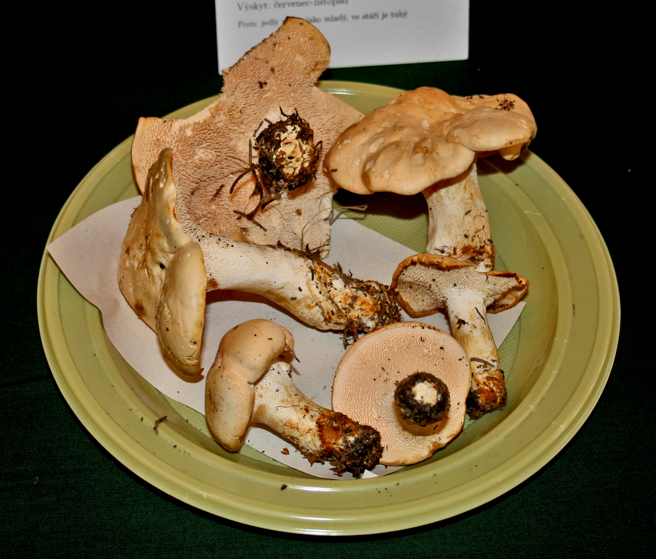


Hydnum repandum, commonly known as the wood hedgehog or hedgehog mushroom, is a distinctive terrestrial fungus characterized by soft, spine-like projections beneath its cap instead of gills or pores. The pale cream to buff-orange cap is firm and fleshy. Widespread across temperate woodlands, it forms associations with both broadleaf and coniferous trees. Renowned for its firm texture and pleasant culinary qualities, it is a staple forest species widely sought across Europe.
Identification, habitat, and key differences.
Forager
Fruitbody structure
- Fruit body form
- Fruit body form: Cap.
Fruit body form: stipe.
Cap
- Cap shape
- in young fruit bodies: Cap shape: convex.
in mature fruit bodies: Cap shape: flat.
Cap shape: depressed. - Cap surface
- Cap surface: dry.
- Cap surface relief
- Cap surface relief: smooth.
Cap surface relief: pitted. - Cap covering
- in young fruit bodies: Cap covering: suede-like.
in old fruit bodies: Cap covering: scaly.
Cap covering: glabrous. - Cap colour
- Cap colour: cream.
Cap colour: yellow-orange.
Cap colour: beige.
Cap colour: whitish.
Cap colour: orange (light shade).
Cap colour: orange-ochre.
Cap colour: orange. - Margin orientation
- in young fruit bodies: Margin orientation: inrolled.
Margin orientation: curved inward. - Cap margin outline
- in old fruit bodies: Cap margin outline: wavy.
in old fruit bodies: Cap margin outline: lobed.
in mature fruit bodies: Cap margin outline: wavy.
in mature fruit bodies: Cap margin outline: lobed. - Cap diameter
- The cap measures up to 150 mm in diameter.
- Cap centre
- Cap centre: Depressed.
Cap underside
- Hymenophore type
- Hymenophore type: spines.
- Hymenophore attachment to the stipe
- Hymenophore attachment to the stipe: attached to the stipe (spines).
- Decurrence of the hymenophore on the stem
- Decurrence of the hymenophore on the stem: decurrent (spines).
- Hymenophore colour
- Hymenophore colour: cream.
Hymenophore colour: yellow-orange.
Hymenophore colour: orange (light shade).
Stem and base
- Stipe position
- Stipe position: off-centre.
- Stipe shape
- Stipe shape: cylindrical (irregular outline).
Stipe shape: club-shaped (slightly). - Stipe colour
- Stipe colour: whitish.
Stipe colour: cream.
Stipe colour: white.
Stipe colour: light brown. - Stipe surface relief
- Stipe surface relief: smooth.
- Stipe interior
- Stipe interior: solid.
- Color of stem surface upon damage
- Color of stem surface upon damage: ochre brown (stipe surface).
Color of stem surface upon damage: no colour change (stipe surface). - Stipe length
- Stipe length: up to 70 mm.
- Stipe diameter
- Stipe diameter: up to 40 mm.
in the upper part of the stipe: Stipe diameter: 7–14 mm. - Stem surface moisture
- Stem surface moisture: dry.
Flesh
- Flesh colour
- Flesh colour: white.
Flesh colour: yellowish.
Flesh colour: whitish. - Flesh firmness
- Flesh firmness: firm.
- Flesh taste
- Flesh taste: sour, weak.
in old fruit bodies: Flesh taste: bitter.
in old fruit bodies: Flesh taste: pungent, weak.
in old fruit bodies: Flesh taste: sour.
Flesh taste: sweet. - Flesh colour after damage
- Flesh colour after damage: no colour change.
- Flesh odour
- Flesh odour: not distinctive.
Flesh odour: somewhat sweet, not distinctive. - Taste character of the flesh
- Taste character of the flesh: mild.
- Distinctiveness of flesh taste
- Distinctiveness of flesh taste: indistinctive.
Where and when it grows
- Habitat
- Habitat: deciduous woodland.
Habitat: coniferous woodland.
Habitat: mixed woodland. - Substrate
- Substrate: soil.
- Fruit body growth pattern
- Fruit body growth pattern: in groups.
Fruit body growth pattern: solitary.
Fruit body growth pattern: in rows.
Fruit body growth pattern: in rings.
Fruit body growth pattern: fused fruit bodies. - Occurrence frequency
- Occurrence frequency: very common.
- Fruiting season
- Fruit bodies appear from July to November.
- Distribution area
- Potwierdzono występowanie w Polsce.
Edibility and culinary use
- Edibility
- Edible
- Required processing
- Requires preparation
Law
- Protection
- It is not subject to species protection in Poland.
- Trade
- It is approved in Poland for trade and for the production of mushroom products.
Lookalikes
Cantharellus cibarius
Key differences
Wood hedgehog (Hydnum repandum) may resemble a chanterelle from above, but under the cap it possesses distinct, dense, brittle spines instead of decurrent folds.
Golden chanterelle (Cantharellus cibarius), commonly called kurka, may resemble the wood hedgehog in coloration and the fleshy structure of the fruitbody, and often grows in the same locations. It differs in the structure of the underside of the cap: the chanterelle has ridges and thick, branched folds, whereas the hedgehog mushroom has distinct, brittle spines.
Hydnum rufescens
Key differences
Terracotta hedgehog (Hydnum rufescens) is characterized by a more intense orange-rufous color and a more slender stem. Discrepancies exist in the literature regarding the attachment of the hymenophore: Snowarski and the ČMS atlas indicate that the spines of the terracotta hedgehog do not run down the stem, whereas Zarzyński gives the opposite trait – more strongly decurrent spines than in the wood hedgehog. Decurrence of spines alone therefore does not constitute an unambiguous distinction criterion across all treatments.
Wood hedgehog (Hydnum repandum) generally differs in larger and paler fruitbodies, a stouter stem merging gently into the cap, and more distinctly decurrent spines. According to AMINT data, fruitbodies of H. repandum frequently grow fused together, which occurs rarely in H. rufescens. The different description of the degree of spine decurrence in Zarzyński's publication remains an isolated discrepancy.
Sarcodon imbricatus
Key differences
The scaly hedgehog (Sarcodon imbricatus) produces distinctly larger fruitbodies, dark grey-brown in colour, covered on the cap surface with large, protruding scales. Although the vernacular name “sarniak” is colloquially used for the wood hedgehog, they are two distinct taxa.
FAQ
What physical feature gives Hydnum repandum its common name of hedgehog mushroom?
The underside of its cap bears soft, tooth-like spines rather than standard gills or pores, resembling the spines of a miniature hedgehog.
Can Hydnum repandum be eaten safely in its raw state?
No, it should not be consumed raw. Thorough cooking is required to prevent digestive discomfort and to neutralize mild gastrointestinal irritants.
Why do cooks sometimes remove the spines from mature specimens?
On older fruiting bodies, the spines can turn bitter and fragile, so removing them before preparation helps preserve a mild, pleasant flavor.
With which types of trees does Hydnum repandum typically form associations?
It associates with various temperate trees, including deciduous species like beech, oak, and chestnut, as well as conifers such as pine, fir, and spruce.
In which regions of the world does Hydnum repandum naturally occur?
In the strict sense, it is verified from Europe, distinguishing it from North American and Japanese sister species, though European verification does not prove absence in all unstudied regions.
What is the typical fruiting season for this species in temperate Europe?
Fruiting generally starts in late summer and continues through late autumn, often extending into early winter in regions with mild maritime climates.
What does the French name pied-de-mouton mean in English?
Pied-de-mouton translates literally to sheep's foot, an evocative vernacular name referring to the shape and texture of the mushroom's cap.
Who provided the formal scientific description of Hydnum repandum?
Swedish botanist Carl Linnaeus formally named and described Hydnum repandum in his landmark 1753 botanical work, Species Plantarum.
How is Hydnum repandum utilized in non-culinary textile crafts?
Dyers boil the fruiting bodies to dye wool and silk, producing subtle cream, golden-yellow, tan, and olive-brown shades depending on the mordant used.
How are preserved specimens of Hydnum repandum used in educational settings?
Natural history museums and universities use dried specimens to teach students and naturalists about non-gilled fungal anatomy.
Enthusiast
Names and history
- Current national name
- Kolczak obłączasty
- Current taxonomic name
- Hydnum repandum
Home observations
Spore print
- Required materials
- fresh, mature cap, sheet of paper, glass or small bowl, water
- Time
- 2–24 h
- Procedure
- Place the cap underside down on the paper.
- Add a drop of water on top and cover.
- After 2–24 hours, lift the cap and examine the deposit.
What to look for
- Spore print colour
- Spore print colour: white.
- Spore print shade
- Spore print shade: pale cream.
Under the microscope
- Required materials
- light microscope, microscope slide and cover slip, needle, water, spores from print
- Procedure
- Transfer a small amount of the spore print onto the slide using a needle.
- Add a drop of water, cover with a cover slip, and examine the spores.
What to look for
- Spore shape
- Spore shape: globose. Spore shape: ovoid (broad). Spore shape: ovoid (broad). Spore shape: globose (nearly). Spore shape: subglobose. Spore shape: broadly ellipsoid. Spore shape: subglobose.
- Spore surface
- Spore surface: smooth. Spore surface: smooth. Spore surface: smooth.
- Spore dimensions
- Length 6–9 µm, width 5–7.5 µm.
- Spore dimensions
- Length 7.8 µm, width 6.8 µm.
Compare dimensions after calibrating the microscope scale.
Ecology and distribution
- Soil reaction
- Soil reaction: alkaline.
- Soil texture
- Soil texture: clayey.
- Soil calcareousness
- Soil calcareousness: calcareous.
- Mycorrhizal hosts
- General species association: Mycorrhizal hosts: Picea (general partner list).
General species association: Mycorrhizal hosts: Fagus (general partner list). - Mode of nutrition
- Mode of nutrition: ectomycorrhizal.
Non-culinary uses
Beyond culinary consumption, Hydnum repandum has established contemporary and historical applications in natural textile dyeing and mycological education.
In fiber arts and botanical dyeing, the species serves as a natural pigment source for coloring protein fibers such as wool and silk. Craft dyers simmer fresh or dried fruiting bodies to extract soluble pigments for textile baths. Unmordanted wool absorbs subtle cream and soft yellowish tones, whereas pre-treating fibers with an alum mordant produces warm golden-yellow and tan hues. When modified with an iron mordant bath, the dye liquor yields muted khaki, olive-brown, and earthy bronze shades. These traditional techniques remain actively practiced by artisan spinners, textile conservators, and historical craft practitioners in Scandinavia and North America seeking plant- and fungal-derived color palettes.
Additionally, the fungus is widely utilized as a physical teaching specimen in educational exhibitions, nature centers, and university mycological programs. Because of its conspicuous spine-bearing underbelly, preserved fruiting bodies and dried herbarium specimens are used to demonstrate non-gilled hymenophore structures to forestry students, naturalists, and the public. Curators also prepare dried reference collections for biodiversity repositories and botanical museum displays.
History and culture
Hydnum repandum holds an enduring place in European folk tradition and botanical history. Carl Linnaeus formally described the species in his 1753 work, Species Plantarum, classifying it among the earliest recognized toothed fungi. Long before binomial nomenclature, agrarian and woodland communities throughout continental Europe recognized the mushroom through vivid vernacular names inspired by its physical characteristics.
In Francophone regions, the mushroom acquired the designation pied-de-mouton (sheep's foot), reflecting folk observations of its pale cap contours. Italian traditions recorded it under names like steccherino dorato (golden pricker), while German-speaking areas established the name Semmel-Stoppelpilz, likening the cap to a pale bread roll (Semmel) paired with bristle-like spines. English folklore solidified the name hedgehog mushroom, drawing on animal imagery to characterize its textured underside.
Beyond vernacular traditions, the species holds documented status in historical European commerce. In nineteenth-century France and Italy, municipal market authorities officially listed the mushroom among approved wild forest commodities authorized for urban trade. This regulatory recognition reflects centuries of established cultural legitimacy and communal valuation across continental Europe.
Edibility and safety
Hydnum repandum is rated as a choice edible mushroom that is consumed across numerous cultures. Safe consumption requires thorough heat treatment; raw or undercooked specimens can trigger gastrointestinal discomfort in sensitive individuals. As fruiting bodies mature, they tend to accumulate a sharp, bitter flavor and a tougher consistency. For older specimens, removing the fragile spine-like structures from the underside of the cap before cooking is a standard practice to reduce bitterness.
Culinary appreciation varies regionally. In France, where it is known as pied-de-mouton, the fungus is highly esteemed and regularly commercialized in traditional produce markets. Italian and Spanish consumers also place high culinary value on young, firm specimens, valuing their slightly peppery taste and capacity to retain firmness when cooked. In Central and Eastern European countries, such as Poland, the Czech Republic, and Germany, it is frequently gathered for home consumption and winter preservation through drying or pickling. Conversely, in parts of North America and the British Isles, consumer enthusiasm has historically been more localized, though appreciation has expanded among wild-food enthusiasts seeking dense-fleshed forest fungi.
Habitat and occurrence
Hydnum repandum in the strict sense is verified from Europe, distinguished from North American sister species like Hydnum neorepandum and Japanese Hydnum repando-orientale, though verification in Europe does not prove its absence in all unstudied regions. The species inhabits mature deciduous, coniferous, and mixed woodland environments, demonstrating a preference for established forest ecosystems with undisturbed floor layers.
Substrate conditions typically consist of moss-covered ground, deep leaf litter, or humus-rich mineral soil, showing a preference for basic soils. In deciduous habitats, the fungus predominantly associates with trees such as European beech (Fagus sylvatica), oaks (Quercus spp.), and sweet chestnut (Castanea sativa). In coniferous stands, it develops alongside Scots pine (Pinus sylvestris), Norway spruce (Picea abies), and silver fir (Abies alba).
The fruiting season spans from late summer through late autumn across northern and central Europe, typically peaking between August and November. In regions with milder maritime climates, such as parts of western Europe, fruitbodies frequently persist into December or January. Conversely, populations in high-elevation montane habitats or continental interiors conclude their fruiting earlier in the autumn before sustained ground freezes arrive.
Sources
- Opracowanie zakresu taksonu oparte na dokładnym dopasowaniu GBIF oraz Niskanen, T. i in. (2018), Identifying and naming the currently known diversity of the genus Hydnum with an emphasis on European and North American taxa, Mycologia 110(5): 890–918, DOI 10.1080/00275514.2018.1477004.
- Zarzyński, P. (2021). Grzyby polskich lasów. Centrum Informacyjne Lasów Państwowych, hasło „Kolczak obłączasty”, drukowana s. 190.
- Tejkal, K. (b.d.). Lišák zprohýbaný (Hydnum repandum L.). Česká mykologická společnost, Myko atlas. https://www.myko.cz/myko-atlas/Hydnum-repandum/
- Kuo, M. (2020, wrzesień). Hydnum repandum. MushroomExpert.Com. https://www.mushroomexpert.com/hydnum_repandum.html
- Snowarski, M. (b.d.). Kolczak obłączasty (Hydnum repandum). Grzyby.pl. https://www.grzyby.pl/gatunki/Hydnum_repandum.htm
Researcher
Edibility and culinary use
Source findings: edibility, consumption, and use
- Edibility
- Edible — identification source
Snowarski, M. (b.d.). Kolczak obłączasty (Hydnum repandum). Grzyby.pl. https://www.grzyby.pl/gatunki/Hydnum_repandum.htm · Edible or poisonous?:Edible.
- Required processing
- Requires preparation
Marttaliitto. Hydnoid fungi: Preparation. · Preparation: hydnoid fungi, young fruitbodies; fry and cook thoroughly.
Scientific names and taxonomy
- Current scientific name
Hydnum repandum
Scientific names and taxonomy
The wood hedgehog (Hydnum repandum L.) was scientifically described by Carl Linnaeus in 1753 in the second volume of the work "Species plantarum" (p. 1178). According to the Species Fungorum database, it belongs to the hydnoid fungus family (Hydnaceae), order Cantharellales (Cantharellales), class Agaricomycetes (Agaricomycetes), division Basidiomycota (Basidiomycota), and kingdom Fungi (Fungi). In the taxonomic revision by Niskanen and co-workers, it constitutes the type species of the genus Hydnum, as well as of the subgenus and section bearing the same name.
Scientific names and taxonomy
In the Species Fungorum register, 27 synonymic entries are recorded for this taxon:
- Hypothele repanda (L.) Paulet, Icon. Champ. (Paris): tab. 35:1-2 (1812)
- Dentinum repandum (L.) Gray, Nat. Arr. Brit. Pl. (London) 1: 650 (1821)
- Tyrodon repandus (L.) P. Karst., Rev. Mycol., Toulouse 3(no. 9): 19 (1881)
- Sarcodon repandus (L.) Quél., Enchiridion Fungorum in Europa media et praesertim in Gallia Vigentium: 189 (1886)
- Hydnum flavidum Schaeff., Fung. bavar. palat. nasc. (Ratisbonae) 4: 99 (1774)
- Hydnum rufescens Schaeff., Fung. bavar. palat. nasc. (Ratisbonae) 4: 95 (1774)
- Sarcodon repandus var. rufescens (Schaeff.) Quél., Enchiridion Fungorum in Europa media et praesertim in Gallia Vigentium: 189 (1886)
- Sarcodon rufescens (Schaeff.) Quél., Fl. mycol. France (Paris): 447 (1888)
- Hydnum clandestinum Batsch, Elench. fung. (Halle): 113 (1783)
- Hydnum repandum var. clandestinum (Batsch) J. Kickx f., in Kickx, Fl. Crypt. Flandres (Paris) 1: 251 (1867)
- Hydnum medium Pers., Observ. mycol. (Lipsiae) 2: 97 (1800) [1799]
- Lophium medium (Pers.) Fr., K. svenska Vetensk-Akad. Handl., ser. 3 39: 113 (1818)
- Hydnum bulbosum Raddi, Mem. Mat. Fis. Soc. Ital. Sci. Modena, Pt. Mem. Fis. 13: 353 (1807)
- Hydnum bicolor Raddi, Mem. Mat. Fis. Soc. Ital. Sci. Modena, Pt. Mem. Fis. 13: 353 (1807)
- Hydnum aurantium Raf., J. Bot. (Desvaux) 1(5): 237 (1813)
- Hydnum repandum var. albidum Fr., Observ. mycol. (Havniae) 1: 139 (1815)
- Hydnum repandum var. denudatum Fr., Observ. mycol. (Havniae) 1: 138 (1815)
- Hydnum repandum var. flavidum Fr., Observ. mycol. (Havniae) 1: 138 (1815)
- Hydnum repandum var. intermedium Fr., Observ. mycol. (Havniae) 1: 139 (1815)
- Hydnum repandum var. rufidum Fr., Observ. mycol. (Havniae) 1: 139 (1815)
- Hydnum album Pers., Traité champ. Comest. (Paris): 249 (1818)
- Hydnum diffractum Berk., London J. Bot. 6: 323 (1847)
- Sarcodon repandus var. albus Quél., Fl. mycol. France (Paris): 447 (1888)
- Hydnum repandum var. album (Quél.) Rea, Brit. basidiomyc. (Cambridge): 630 (1922)
- Dentinum repandum var. album (Quél.) K.A. Harrison, Publs Dep. Agric. Can. 1099: 19 (1961)
- Hydnum washingtonianum Ellis & Everh., Proc. Acad. nat. Sci. Philad. 46(3): 323 (1894)
- Hydnum repandum var. albidum Cejp, Fauna Flora Cechoslov., II, Hydnaceae: 82 (1928)
This compilation documents the history of name usage in mycological literature, including, among others, two distinct treatments of variety albidum and the dating of Hydnum medium from 1800 [1799].
Scientific names and taxonomy
In historical mycological literature, this species was transferred to other genera, creating the combinations Dentinum repandum, Hypothele repanda, Tyrodon repandus, and Sarcodon repandus. The synonymy also includes Hydnum rufescens Schaeff. from 1774, whose authorship is significant because it is not identical to Hydnum rufescens Pers. from 1799 (recognized in the 2018 revision as a distinct taxon — the terracotta hedgehog). Furthermore, in Snowarski's Polish atlas, the historical form Hydnum repandum Fr. f. minor Nikol. was noted.
- Zakres ujęcia taksonomicznego
Hydnum repandum L. w dokładnym zaakceptowanym dopasowaniu GBIF2554716 oraz europejskim ujęciu rewizji Niskanen i in.2018. Historyczne opisy szerszego kompleksu, wzmianki północnoamerykańskie i etykiety sekwencji pozostają przy własnych źródłach; nie dostarczają automatycznie obecnemu gatunkowi zasięgu lub cech jego sobowtórów.
[2] [3]
Ecology and distribution
- Occurrence frequency
- Poland: Occurrence frequency: very common. [18]
Laboratory diagnostics
Structured data
Basidia
- Clamp connections
- Clamp connections: present. [16]
Hymenial cystidia
- Presence of hymenial pseudocystidia
- Presence of hymenial pseudocystidia: present. [16]
- Shape of hymenial pseudocystidia
- Shape of hymenial pseudocystidia: cylindrical (mostly). [16]
- Width of hymenial pseudocystidia
- Width of hymenial pseudocystidia: 3–8 µm. [16]
- Length of hymenial pseudocystidia
- Length of hymenial pseudocystidia: 30–40 µm. [16]
Other laboratory characteristics
Pileipellis
- Optical appearance of pileipellis hyphae
- Optical appearance of pileipellis hyphae: hyaline. [16]
- Arrangement of pileipellis hyphae
- Arrangement of pileipellis hyphae: interwoven. [16]
- Width of pileipellis elements
- Width of pileipellis elements: 2–6 µm. [16]
- Colour of pileipellis hyphae
- Colour of pileipellis hyphae: ochre-coloured. [16]
Spores
- Mean spore length-to-width ratio
- Mean spore length-to-width ratio: 1.15 (mean). [3]
- Spore colour under the microscope
- Spore colour under the microscope: hyaline. [18]
Spore colour under the microscope: hyaline. [16] - Spore length
- Spore length: 6.5–9 µm. [18]
Spore length: 7.8 µm (mean). [3]
Spore length: 7–9 µm. [9]
Spore length: 7–8.5 µm. [3]
Spore length: 6–8 µm. [16] - Spore length-to-width ratio
- Spore length-to-width ratio: 1.08–1.25. [3]
- Spore shape
- Spore shape: globose. [18]
Spore shape: ovoid (broad). [18]
Spore shape: ovoid (broad). [9]
Spore shape: globose (nearly). [9]
Spore shape: subglobose. [16]
Spore shape: broadly ellipsoid. [16]
Spore shape: subglobose. [3] - Spore wall thickness
- Spore wall thickness: thin-walled. [3]
Material and research methods
Description of characteristics — source and scope
Material: Source species description; without own specimen and without default protocol.
Reading scope: Full text.
Microscopic data — source and limitations
Material: Source species description; without own specimen and without default protocol.
Reading scope: Full text.
Study of composition and activity — Çayan F (2021)
Material: Dried fruit bodies of H. repandum collected in Fethiye, Muğla Province, Turkey, in 2018; the material was ground to particles of approx. 0,5 mm in size. The article states that specimens with voucher numbers were deposited in the herbarium of Muğla Sıtkı Koçman University, but the voucher number of H. repandum was not indicated in the read text.
Reading scope: Full text.
Limitations: In this revision, only results for H. repandum were taken into account; data of the other four species were not assigned to this taxon. The exact voucher number of H. repandum does not appear in the read material description. Table 1 provides values with a ± sign, but does not specify whether they are means nor whether the uncertainty is SD or SEM. The reported value and unspecified type of ± were retained.
Study of composition and activity — Rašeta M
Material: Wild fruit bodies of H. repandum were collected in January 2021 on Mount Tara, in the town of Mitrovac in Serbia. The identification was made by Dr. Eleonora Čapelja from the Profungi laboratory at the University of Novi Sad; the voucher was deposited in the herbarium of this university under the number BUNS 12-00710.
Reading scope: Source fragments.
A fragment of the source was read; full text and methods were not verified.
Material preparation: Extract with 70% ethanol from 10 g of material H. repandum; the source does not state the volume of ethanol used for maceration. 1. extraction — ethanol.
Material preparation: Extract with 80% methanol from 10 g of material H. repandum using 100 mL of solvent. 1. extraction — methanol.
Limitations: The preserved Figshare file is a PDF supplement; it is not a separately downloaded publisher full text. Measurements come from a single collection of fruit bodies; sample size of the biological sample was not given. The caption of Figure S1 specifies the data as mean ± SD and provides Tukey's HSD test; the number of replicates was not indicated in the preserved material. In this compilation, values for 70% EtOH and 80% MeOH extracts mentioned in the abstract were included; values from the graphs were not reconstructed. Results from the supplement table were preserved in a separate compilation.
Study of composition and activity — Çayan F (2021)
Material: Hexane and methanol extracts from dried, powdered fruit bodies of H. repandum; Fethiye, Turkey, 2018.
Reading scope: Full text.
Limitations: For the boundaries >0,50 mg/mL, neither numerical uncertainty nor the number of independent biological samples was provided.
- Results as recorded in the sources — Spores per basidium
- Liczba zarodników na jednej podstawce wynosi 4. [3] [16]
- Results as recorded in the sources — Spore length
- 7–8,5 µm [3]
6–8 µm [16]
6,5–9 µm [18]
Materiał badania: Ujęcie taksonomiczne Hydnum repandum w pracy Niskanen i in. (2018); praca wyznacza epityp z Finlandii i zawiera diagnozę gatunku opartą na omawianym materiale taksonomicznym. Z kolców każdego owocnika zmierzono 20 zarodników, pomijając najmniejsze. Średnia długość zarodnika w badanym materiale wynosi 7,8 mikrometra. [3]
7–9 µm [9] - Results as recorded in the sources — Spore width
- Materiał badania: Ujęcie taksonomiczne Hydnum repandum w pracy Niskanen i in. (2018); praca wyznacza epityp z Finlandii i zawiera diagnozę gatunku opartą na omawianym materiale taksonomicznym. Z kolców każdego owocnika zmierzono 20 zarodników, pomijając najmniejsze. Średnia szerokość zarodnika w badanym materiale wynosi 6,8 mikrometra. [3]
5–7,5 µm [9]
5,5–7 µm [18] [16]
6,2–7,5 µm [3]
Metody i zapisy źródłowe — Opis Hydnum repandum Michaela Kuo w MushroomExpert.Com, wersja z września 2020 r.; obejmuje osobne noty o rozmiarach struktur i ich wyglądzie w KOH.
Metoda źródłowa. Technika: mikroskopia świetlna (LM). Medium: KOH. Opis metody w źródle: Źródło podaje KOH; stężenia nie podano.
Wygląd optyczny strzępek skórki — zapis źródłowy: „Cap surface hyphae hyaline in KOH.” [16]
Układ strzępek skórki — zapis źródłowy: „Surface hyphae tangled; pileipellis not clearly differentiated.” [16]
Obecność pseudocystyd hymenialnych — zapis źródłowy: „Pseudocystidia 30–40 × 3–8 µm.” [16]
Kształt pseudocystyd hymenialnych — zapis źródłowy: „Mostly more or less cylindric.” [16]
Szerokość pseudocystyd hymenialnych — zapis źródłowy: „Pseudocystidia 3–8 µm wide.” [16]
Liczba zarodników na jednej podstawce — zapis źródłowy: „Basidia 4-sterigmate.” [16]
Długość podstawki; Szerokość podstawki — zapis źródłowy: „Basidia 29–34 × 5–6 µm.” [16]
Kształt podstawki — zapis źródłowy: „Basidia subclavate.” [16]
Długość pseudocystyd hymenialnych — zapis źródłowy: „Pseudocystidia 30–40 µm long.” [16]
Szerokość elementów skórki — zapis źródłowy: „Surface hyphae 2–6 µm wide.” [16]
Barwa strzępek skórki kapelusza — zapis źródłowy: „Cap surface hyphae ochraceous in KOH.” [16]
Barwa zarodnika w mikroskopie — zapis źródłowy: „Spores hyaline in KOH.” [16]
Długość zarodnika; Kształt zarodnika; Powierzchnia zarodnika; Szerokość zarodnika — zapis źródłowy: „Microscopic Features: source sizes and separate KOH colour context; no medium attached to shape or measurements.” [16]
Metody i zapisy źródłowe — Ujęcie taksonomiczne Hydnum repandum w pracy Niskanen i in. (2018); praca wyznacza epityp z Finlandii i zawiera diagnozę gatunku opartą na omawianym materiale taksonomicznym. Z kolców każdego owocnika zmierzono 20 zarodników, pomijając najmniejsze.
Długość sterygmy podstawki — zapis źródłowy: „Sterigmata 3–6 µm; not spore appendage.” [3]
Liczba zarodników na jednej podstawce — zapis źródłowy: „Basidia 4-spored.” [3]
Długość podstawki; Szerokość podstawki — zapis źródłowy: „Basidia 35–45 × 6–8 µm.” [3]
Długość zarodnika; Kształt zarodnika; Szerokość zarodnika — zapis źródłowy: „Hydnum repandum species description: PDF p21–22; spores 7–8.5 × 6.2–7.5 µm, Q 1.08–1.25; basidia 35–45 × 6–8 µm, 4-spored, sterigmata 3–6 µm.” [3]
Długość zarodnika — zapis źródłowy: „Reported average spore length 7.8 µm; not range extrema.” [3]
Iloraz długości i szerokości zarodnika — zapis źródłowy: „Reported Q 1.08–1.25; not derived from independent extrema.” [3]
Grubość ściany zarodnika — zapis źródłowy: „Spores thin-walled.” [3]
Szerokość zarodnika — zapis źródłowy: „Reported average spore width 6.8 µm; not range extrema.” [3]
Metody i zapisy źródłowe — Opis mikroskopowy Hydnum repandum przytoczony przez Marka Snowarskiego na stronie Grzyby.pl; ujęcie atlasowe, a nie własny pomiar zapisany w tym dokumencie.
Barwa zarodnika w mikroskopie — zapis źródłowy: „Bezbarwne; medium niepodane.” [18]
Długość zarodnika; Kształt zarodnika; Powierzchnia zarodnika; Szerokość zarodnika — zapis źródłowy: „Zarodniki: 6,5–9 × 5,5–7 µm; kuliste do szeroko jajowatych, gładkie, bezbarwne.” [18]
Metody i zapisy źródłowe — Mikroskopowy opis Hydnum repandum w atlasie Myko České mykologické společnosti, w sekcji „Výtrusy”; stronę opracował Karel Tejkal.
Długość zarodnika; Kształt zarodnika; Powierzchnia zarodnika; Szerokość zarodnika — zapis źródłowy: „Popis: výtrusy 7–9 × 5–7,5 µm, široce oválné až téměř kulovité, hladké.” [9]
Occurrence verification — scope and limitations
Material: Epityp H6003710 z Ruissalo, Turku; pojedyncza lokalizacja.
Reading scope: Full text.
Review date: 30.10.2026.
Limitations: One collection does not establish the full range of Finland.
Pure mycelial culture — material and limitations
Material: A pure mycelial culture was obtained from fruitbodies collected in the autumn of 2008 in a pine forest in Sinop province, Turkey.
Reading scope: Full text.
Limitations: The authors describe the identification as conventional, according to Phillips (1994); they do not provide a voucher number, DNA sequence, strain identifier, or accession. The results concern controlled mycelial growth and vegetative inoculum; they are not a measurement of natural fruitbody mass or fruitbody yield. Tables 1–3 report means and Duncan test grouping letters, without SD or SEM. Table 4 designates the results as ns; it does not report SD or SEM.
Pure mycelial culture — results report
Culture conditions: In darkness, on day 18 of culture. Results under these conditions:
Table 4 — mean mycelial growth rate reported in the source as 1.08 cm/day (= 10.8 mm/day) in peat. Inoculum in a 1 L bottle: 800 mL of substrate moistened with 200 mL of liquid MEPA; four mycelial discs with a diameter of 5 mm; 20±2 °C, darkness; n=4 bottles per variant. Growth was measured daily in two parts; growth length was reported for day 18. No SD/SEM; variant effects in the table were marked ns.
Table 4 — mean mycelial growth rate reported in the source as 1.07 cm/day (= 10.7 mm/day) in a peat:vermiculite mixture 1:4 (v:v). Inoculum in a 1 L bottle: 800 mL of substrate moistened with 200 mL of liquid MEPA; four mycelial discs with a diameter of 5 mm; 20±2 °C, darkness; n=4 bottles per variant. Growth was measured daily in two parts; growth length was reported for day 18. No SD/SEM; variant effects in the table were marked ns.
Table 4 — mean mycelial growth rate reported in the source as 1.06 cm/day (= 10.6 mm/day) in a peat:vermiculite mixture 1:6 (v:v). Inoculum in a 1 L bottle: 800 mL of substrate moistened with 200 mL of liquid MEPA; four mycelial discs with a diameter of 5 mm; 20±2 °C, darkness; n=4 bottles per variant. Growth was measured daily in two parts; growth length was reported for day 18. No SD/SEM; variant effects in the table were marked ns.
Table 4 — mean mycelial growth rate reported in the source as 1.14 cm/day (= 11.4 mm/day) in a peat:vermiculite mixture 1:8 (v:v). Inoculum in a 1 L bottle: 800 mL of substrate moistened with 200 mL of liquid MEPA; four mycelial discs with a diameter of 5 mm; 20±2 °C, darkness; n=4 bottles per variant. Growth was measured daily in two parts; growth length was reported for day 18. No SD/SEM; variant effects in the table were marked ns.
Table 4 — mean mycelial growth rate reported in the source as 0.94 cm/day (= 9.4 mm/day) in a peat:vermiculite mixture 1:10 (v:v). Inoculum in a 1 L bottle: 800 mL of substrate moistened with 200 mL of liquid MEPA; four mycelial discs with a diameter of 5 mm; 20±2 °C, darkness; n=4 bottles per variant. Growth was measured daily in two parts; growth length was reported for day 18. No SD/SEM; variant effects in the table were marked ns.
Culture conditions: At a temperature of 15 degrees Celsius, in darkness, on day 15 of culture. Results under these conditions:
Table 1 — mean colony diameter: 58.98 mm after 15 days, at 15 °C and in darkness, on PDYA; mean for six initial pH values: 4.0, 4.5, 5.0, 5.5, 6.0 and 6.5; 5 plates for each pH and temperature combination. The table does not report SD or SEM.
Culture conditions: At a temperature of 20 degrees Celsius, in darkness, on day 15 of culture. Results under these conditions:
Table 1 — mean colony diameter: 80.10 mm after 15 days, at 20 °C and in darkness, on PDYA; mean for six initial pH values: 4.0, 4.5, 5.0, 5.5, 6.0 and 6.5; 5 plates for each pH and temperature combination. The table does not report SD or SEM.
Table 1 — mean colony diameter: 86.18 mm after 15 days, on PDYA, at initial pH 5.5 and temperature 20 °C, in darkness; n=5 plates for this combination. The table does not report SD or SEM.
Culture conditions: At a temperature of 25 degrees Celsius, in darkness, on day 15 of culture. Results under these conditions:
Table 1 — mean colony diameter: 76.88 mm after 15 days, at 25 °C and in darkness, on PDYA; mean for six initial pH values: 4.0, 4.5, 5.0, 5.5, 6.0 and 6.5; 5 plates for each pH and temperature combination. The table does not report SD or SEM.
Table 1 — mean colony diameter: 88.21 mm after 15 days, on PDYA, at initial pH 5.5 and temperature 25 °C, in darkness; n=5 plates for this combination. The table does not report SD or SEM.
Culture conditions: At a temperature of 25 degrees Celsius, in darkness, on day 11 of culture. Results under these conditions:
Table 2 — mean colony diameter after 11 days: 53.98 mm; PDYA medium with added xylose (xylose) at a concentration of 20 g/L; 25 °C, darkness; n=9 plates per variant. The table reports means and Duncan's grouping letters; SD/SEM not reported.
Table 2 — mean colony diameter after 11 days: 59.26 mm; PDYA medium with added lactose (lactose) at a concentration of 20 g/L; 25 °C, darkness; n=9 plates per variant. The table reports means and Duncan's grouping letters; SD/SEM not reported.
Table 2 — mean colony diameter after 11 days: 52.79 mm; PDYA medium with added sucrose (sucrose) at a concentration of 20 g/L; 25 °C, darkness; n=9 plates per variant. The table reports means and Duncan's grouping letters; SD/SEM not reported.
Table 2 — mean colony diameter after 11 days: 68.05 mm; PDYA medium with added maltose (maltose) at a concentration of 20 g/L; 25 °C, darkness; n=9 plates per variant. The table reports means and Duncan's grouping letters; SD/SEM not reported.
Table 2 — mean colony diameter after 11 days: 82.72 mm; PDYA medium with added mannitol (mannitol) at a concentration of 20 g/L; 25 °C, darkness; n=9 plates per variant. The table reports means and Duncan's grouping letters; SD/SEM not reported.
Table 2 — mean colony diameter after 11 days: 81.65 mm; PDYA medium with added glucose (glucose) at a concentration of 20 g/L; 25 °C, darkness; n=9 plates per variant. The table reports means and Duncan's grouping letters; SD/SEM not reported.
Table 2 — mean colony diameter after 11 days: 73.15 mm; PDYA medium with added dextrose (dextrose) at a concentration of 20 g/L; 25 °C, darkness; n=9 plates per variant. The table reports means and Duncan's grouping letters; SD/SEM not reported.
Table 2 — mean colony diameter after 11 days: 48.87 mm; PDYA medium, control without a carbon source (C-free control); 25 °C, darkness; n=9 plates per variant. The table reports the mean and Duncan's grouping letters; SD/SEM not reported.
Table 3 — mean colony diameter after 11 days: 56.63 mm; PDYA medium with added (NH4)2HPO4 at a concentration of 2 g/L; 25 °C, darkness; n=9 plates per variant. The table reports means and Duncan's grouping letters; SD/SEM not reported.
Table 3 — mean colony diameter after 11 days: 66.47 mm; PDYA medium with added NH4NO3 at a concentration of 2 g/L; 25 °C, darkness; n=9 plates per variant. The table reports means and Duncan's grouping letters; SD/SEM not reported.
Table 3 — mean colony diameter after 11 days: 80.27 mm; PDYA medium with added peptone (peptone) at a concentration of 2 g/L; 25 °C, darkness; n=9 plates per variant. The table reports means and Duncan's grouping letters; SD/SEM not reported.
Table 3 — mean colony diameter after 11 days: 86.20 mm; PDYA medium with added Ca(NO3)2 at a concentration of 2 g/L; 25 °C, darkness; n=9 plates per variant. The table reports means and Duncan's grouping letters; SD/SEM not reported.
Table 3 — mean colony diameter after 11 days: 72.59 mm; PDYA medium with added malt extract (malt extract) at a concentration of 2 g/L; 25 °C, darkness; n=9 plates per variant. The table reports means and Duncan's grouping letters; SD/SEM not reported.
Table 3 — mean colony diameter after 11 days: 69.66 mm; PDYA medium with added yeast extract (yeast extract) at a concentration of 2 g/L; 25 °C, darkness; n=9 plates per variant. The table reports means and Duncan's grouping letters; SD/SEM not reported.
Table 3 — mean colony diameter after 11 days: 72.69 mm; PDYA medium, control without a nitrogen source (N-free control); 25 °C, darkness; n=9 plates per variant. The table reports the mean and Duncan's grouping letters; SD/SEM not reported.
Scope of the study
Before completion, the record requires completion or settlement.
- Recorded source information is awaiting resolution.
- New sources require accounting for information encountered during the review.
Information pending resolution: 6
Legal basis and scope of assessment
Regulations for location: Poland
Basis of protection information
The applicable regulation Dz.U. 2014 item 1408 does not list Hydnum repandum or wood hedgehog in the species protection lists of fungi in Annexes 1–4.
Basis of trade information
Hydnum repandum L. figuruje under item 14 of Annex No. 1 to the applicable consolidated text published in Dz.U. 2026 item 258. § 2 para. 1 of this regulation designates Annex No. 1 as the list of mushrooms permitted for marketing or the production of processed products and foodstuffs containing mushrooms.
Analytical results
Composition and laboratory activity
- Quantitative content of component in material
- Quantitative content of component in material: 0.037 mg/100g (4-hydroxybenzoic acid, fruit body preparation, assay replicates, number of replicates: 3, unspecified measure of variation reported as ±, on a dry mass basis, dried fruit-body powder used in the study by Çayan et al., reported value after ±: 0.002, reported value). [1]
Quantitative content of component in material: 0.001 mg/100g (p-coumaric acid, fruit body preparation, assay replicates, number of replicates: 3, unspecified measure of variation reported as ±, on a dry mass basis, dried fruit-body powder used in the study by Çayan et al., reported value after ±: 0.001, reported value). [1]
Quantitative content of component in material: 0.001 mg/100g (ferulic acid, fruit body preparation, assay replicates, number of replicates: 3, unspecified measure of variation reported as ±, on a dry mass basis, dried fruit-body powder used in the study by Çayan et al., reported value after ±: 0.001, reported value). [1]
Quantitative content of component in material: 0.023 mg/100g (gallic acid, fruit body preparation, assay replicates, number of replicates: 3, unspecified measure of variation reported as ±, on a dry mass basis, dried fruit-body powder used in the study by Çayan et al., reported value after ±: 0.001, reported value). [1]
Quantitative content of component in material: 0.08671 mg/g (fumaric acid, fruit body preparation, assay replicates, number of replicates: 3, unspecified measure of variation reported as ±, on a dry mass basis, dried fruit-body powder used in the study by Çayan et al., reported value after ±: 0.00005, reported value). [1]
Quantitative content of component in material: 0.011 mg/100g (caffeic acid, fruit body preparation, assay replicates, number of replicates: 3, unspecified measure of variation reported as ±, on a dry mass basis, dried fruit-body powder used in the study by Çayan et al., reported value after ±: 0.003, reported value). [1] - Reducing capacity in the Folin–Ciocalteu assay
- Reducing capacity in the Folin–Ciocalteu assay: 27.44 mgGAE/g (Folin–Ciocalteu assay, extract, relative to the mass of the starting material, on a dry mass basis, standard deviation: 0.32, extract obtained using 70% ethanol, calibration standard: gallic acid, mean). [7]
Reducing capacity in the Folin–Ciocalteu assay: 28.29 mgGAE/g (Folin–Ciocalteu assay, extract, relative to the mass of the starting material, on a dry mass basis, standard deviation: 3.62, extract obtained using 80% methanol, calibration standard: gallic acid, mean). [7]
Separate source analytical results
- p-hydroxybenzoic acid
- The determination result is less than 3,05 ng/mL. The mass basis was not stated in the source. Method according to source: LC-MS/MS analysis.[7]
- p-hydroxybenzoic acid
- The determination result is 16,84112 ng/mL. The mass basis was not stated in the source. Method according to source: LC-MS/MS analysis.[7]
- p-hydroxybenzoic acid
- The determination result is 6,352015 ng/mL. The mass basis was not stated in the source. Method according to source: LC-MS/MS analysis.[7]
- p-hydroxybenzoic acid
- The determination result is 2,814165 ng/mL. The mass basis was not stated in the source. Method according to source: LC-MS/MS analysis.[7]
- p-hydroxybenzoic acid
- The determination result is 11,98769 ng/mL. The mass basis was not stated in the source. Method according to source: LC-MS/MS analysis.[7]
- protocatechuic acid
- The determination result is less than 1,525 ng/mL. The mass basis was not stated in the source. Method according to source: LC-MS/MS analysis.[7]
- protocatechuic acid
- The determination result is 19,09858 ng/mL. The mass basis was not stated in the source. Method according to source: LC-MS/MS analysis.[7]
- protocatechuic acid
- The determination result is 4,82905 ng/mL. The mass basis was not stated in the source. Method according to source: LC-MS/MS analysis.[7]
- protocatechuic acid
- The determination result is 9,404285 ng/mL. The mass basis was not stated in the source. Method according to source: LC-MS/MS analysis.[7]
- protocatechuic acid
- The determination result is 16,92327 ng/mL. The mass basis was not stated in the source. Method according to source: LC-MS/MS analysis.[7]
- 2,5-dihydroxybenzoic acid
- The determination result is less than 1,525 ng/mL. The mass basis was not stated in the source. Method according to source: LC-MS/MS analysis.[7]
- 2,5-dihydroxybenzoic acid
- The determination result is 5,66008 ng/mL. The mass basis was not stated in the source. Method according to source: LC-MS/MS analysis.[7]
- 2,5-dihydroxybenzoic acid
- The determination result is less than 1,525 ng/mL. The mass basis was not stated in the source. Method according to source: LC-MS/MS analysis.[7]
- 2,5-dihydroxybenzoic acid
- The determination result is 2,340995 ng/mL. The mass basis was not stated in the source. Method according to source: LC-MS/MS analysis.[7]
- 2,5-dihydroxybenzoic acid
- The determination result is 5,743895 ng/mL. The mass basis was not stated in the source. Method according to source: LC-MS/MS analysis.[7]
- quinic acid
- The determination result is less than 1,525 ng/mL. The mass basis was not stated in the source. Method according to source: LC-MS/MS analysis.[7]
- quinic acid
- The determination result is 676,0725 ng/mL. The mass basis was not stated in the source. Method according to source: LC-MS/MS analysis.[7]
- quinic acid
- The determination result is less than 1,525 ng/mL. The mass basis was not stated in the source. Method according to source: LC-MS/MS analysis.[7]
- quinic acid
- The determination result is 648,5988 ng/mL. The mass basis was not stated in the source. Method according to source: LC-MS/MS analysis.[7]
- quinic acid
- The determination result is 531,3731 ng/mL. The mass basis was not stated in the source. Method according to source: LC-MS/MS analysis.[7]
- chrysoeriol
- The determination result is 4,18907 ng/mL. The mass basis was not stated in the source. Method according to source: LC-MS/MS analysis.[7]
- chrysoeriol
- The determination result is 2,16884 ng/mL. The mass basis was not stated in the source. Method according to source: LC-MS/MS analysis.[7]
- chrysoeriol
- The determination result is less than 1,525 ng/mL. The mass basis was not stated in the source. Method according to source: LC-MS/MS analysis.[7]
- chrysoeriol
- The determination result is less than 1,525 ng/mL. The mass basis was not stated in the source. Method according to source: LC-MS/MS analysis.[7]
- chrysoeriol
- The determination result is less than 1,525 ng/mL. The mass basis was not stated in the source. Method according to source: LC-MS/MS analysis.[7]
- amentoflavone
- The determination result is 61,66483 ng/mL. The mass basis was not stated in the source. Method according to source: LC-MS/MS analysis.[7]
- amentoflavone
- The determination result is 34,73948 ng/mL. The mass basis was not stated in the source. Method according to source: LC-MS/MS analysis.[7]
- amentoflavone
- The determination result is 21,629 ng/mL. The mass basis was not stated in the source. Method according to source: LC-MS/MS analysis.[7]
- amentoflavone
- The determination result is 17,14022 ng/mL. The mass basis was not stated in the source. Method according to source: LC-MS/MS analysis.[7]
- amentoflavone
- The determination result is 11,8142 ng/mL. The mass basis was not stated in the source. Method according to source: LC-MS/MS analysis.[7]
Separate source analytical results
- coumarin
- The determination result is 0,82 micrograms per gram. The result refers to the dry weight of the tested material. Uncertainty: 0,09 micrograms per gram (type of uncertainty not specified in the source). n = 3 (replicates stated in the source; biochemical measurements). Method according to source: HPLC-DAD.[1]
- trans-2-hydroksycynamonowy acid
- The determination result is 0,71 micrograms per gram. The result refers to the dry weight of the tested material. Uncertainty: 0,06 micrograms per gram (type of uncertainty not specified in the source). n = 3 (replicates stated in the source; biochemical measurements). Method according to source: HPLC-DAD.[1]
- ellagic acid
- The determination result is 0,31 micrograms per gram. The result refers to the dry weight of the tested material. Uncertainty: 0,04 micrograms per gram (type of uncertainty not specified in the source). n = 3 (replicates stated in the source; biochemical measurements). Method according to source: HPLC-DAD.[1]
- rosmarinic acid
- The determination result is 3,62 micrograms per gram. The result refers to the dry weight of the tested material. Uncertainty: 0,07 micrograms per gram (type of uncertainty not specified in the source). n = 3 (replicates stated in the source; biochemical measurements). Method according to source: HPLC-DAD.[1]
- trans-cinnamic acid
- The determination result is 0,26 micrograms per gram. The result refers to the dry weight of the tested material. Uncertainty: 0,02 micrograms per gram (type of uncertainty not specified in the source). n = 3 (replicates stated in the source; biochemical measurements). Method according to source: HPLC-DAD.[1]
Key substances and their effects
Main substances and their action
In the revision of the genus Hydnum from 2018, Niskanen and coworkers combined morphological characters with DNA sequence analysis of the ITS region, recognizing 49 species worldwide, including 22 newly described. As part of the research, 46 ITS sequences from 18 species were generated, and the final phylogenetic matrix included 132 sequences with a length of 695 aligned positions. The analysis was conducted using the maximum likelihood method in the RAxML 8 program with the GTRGAMMA evolution model and 1000 bootstrap replicates.
Main substances and their action
Genetic material was isolated from dried spines using the NucleoSpin Plant kit, and amplification was performed with primers ITS1F and ITS4. Sequences were assembled in Sequencher 4.1, aligned with the MUSCLE algorithm, and adjusted manually in the SeaView tool. Sequences longer than 400 base pairs were included in the dataset, omitting identical sequences from the same region and most taxa from Australasia. The resulting alignment was deposited in the TreeBASE database under number S21634.
Main substances and their action
In the phylogenetic tree, Hydnum repandum formed a paraphyletic grade with respect to three closely related lineages: H. neorepandum, H. boreorepandum, and H. repando-orientale, which obtained bootstrap support above 90%. Intraspecific variation in this group was below 0,5%, whereas interspecific differences were at a level of about 1%, which, along with morphological and geographical distinctness, served to maintain them as separate species.
Main substances and their action
In ethnomycological research in the Mazovia region, the method of semi-structured interviews and free-listing of known mushrooms was used, verified by herbarium specimens, field walks, and ITS analyses. The traditional group Hydnum repandum sensu lato, also including H. ellipsosporum, obtained four use citations in the study and Smith's cultural salience index S = 0,0046.
Habitat and occurrence
Habitat, area, and season
In the Czech Republic, the species appears quite frequently from July to November. It grows in deciduous and coniferous forests, forming extensive clusters, sometimes arranged in regular rows or fairy rings.
Habitat, area, and season
In the taxonomic revision from 2018, the occurrence of Hydnum repandum sensu stricto was confirmed in Europe from late summer to late autumn. The fungus was recorded in forest stands dominated by Norway spruce (Picea abies) with the presence of birch (Betula), pine (Pinus), poplar (Populus), hazel (Corylus), or oak (Quercus), as well as in fir (Abies) and beech (Fagus) forests. The epitype specimen was collected under Scots pine (Pinus sylvestris) with oaks growing in the vicinity.
Habitat, area, and season
The earlier, broad concept of the species encompassed North America and Europe. However, phylogenetic studies from 2018 demonstrated the presence of distinct species: the North American Hydnum neorepandum and the Japanese Hydnum repando-orientale. In turn, sequence JQ063050 from Venezuela, isolated from mycorrhiza on the roots of Pakaraimaea dipterocarpacea, requires further confirmation due to the unusual geographical range and a different host.
Habitat, area, and season
In the Czech mycological database ČMS, specific field finds were documented: 15 August 2020 in the Central Bohemian Region (Středočeský kraj, 0071-2020-0142), 18 September 2021 in the Plzeň Region (Plzeňský kraj, 0071-2021-0273), 11 September 2022 in Slovakia (0195-2022-0015), and 6 October 2023 in the Zlín Region (Zlínský kraj, 0066-2023-0289).
Confirmed distribution range
All areas confirmed in recorded source data.
Sources
- Çayan F, Tel-Çayan G, Deveci E, Duru ME. HPLC–DAD characterization of phenolic profile and in vitro antioxidant, anticholinesterase, and antidiabetic activities of five mushroom species from Turkey. 3 Biotech. 2021;11(6): 273. DOI: 10.1007/s13205-021-02819-3. PMCID: PMC8128964.
- GBIF Secretariat. Species Match API, dokładne dopasowanie Hydnum repandum, zapytanie z 30.10.2026. https://api.gbif.org/v1/species/match?name=Hydnum%20repandum&strict=true&rank=SPECIES
- Opracowanie zakresu taksonu oparte na dokładnym dopasowaniu GBIF oraz Niskanen, T. i in. (2018), Identifying and naming the currently known diversity of the genus Hydnum with an emphasis on European and North American taxa, Mycologia 110(5): 890–918, DOI 10.1080/00275514.2018.1477004.
- Podstawa: Rozporządzenie Ministra Środowiska z 9 października 2014 r., Dz.U. 2014 poz. 1408, załączniki 1–4; oraz Obwieszczenie Ministra Zdrowia z 13 lutego 2026 r., Dz.U. 2026 poz. 258, załącznik nr 1, lp. 14.
- Peksen A, Kibar B, Yakupoglu G. Favourable Culture Conditions for Mycelial Growth of Hydnum Repandum, a Medicinal Mushroom. African Journal of Traditional, Complementary and Alternative Medicines. 2013;10(6): 431–434. DOI: 10.4314/ajtcam.v10i6.4. PMCID: PMC3847377.
- Rašeta M, Mišković J, Berežni S, Kostić S, Kebert M, Matavulj M, Karaman M. Antioxidant proficiency in Serbian mushrooms: a comparative study on Hydnum repandum L. 1753 from mycorrhizal and edible niches. DOI: 10.1080/14786419.2024.2341300. Figshare DOI: 10.6084/m9.figshare.25579831.v1.
- Tejkal, K. (b.d.). Lišák zprohýbaný (Hydnum repandum L.). Česká mykologická společnost, Myko atlas. https://www.myko.cz/myko-atlas/Hydnum-repandum/ (also [10])
- Rozporządzenie Ministra Środowiska z dnia 9 października 2014 r. w sprawie ochrony gatunkowej grzybów, Dz.U. 2014 poz. 1408, ELI: DU/2014/1408.
- ELI/Sejm RP. Metryka aktu: Rozporządzenie Ministra Środowiska z dnia 9 października 2014 r. w sprawie ochrony gatunkowej grzybów, Dz.U. 2014 poz. 1408; status sprawdzony 30.10.2026.
- Obwieszczenie Ministra Zdrowia z dnia 13 lutego 2026 r. w sprawie ogłoszenia tekstu jednolitego rozporządzenia dotyczącego grzybów dopuszczonych do obrotu lub produkcji przetworów, Dz.U. 2026 poz. 258, załącznik nr 1, lp. 14.
- ELI/Sejm RP. Metryka aktu: Obwieszczenie Ministra Zdrowia z dnia 13 lutego 2026 r., Dz.U. 2026 poz. 258; status sprawdzony 30.10.2026.
- Kotowski, M. A., Pietras, M., & Łuczaj, Ł. (2019). Extreme levels of mycophilia documented in Mazovia, a region of Poland. Journal of Ethnobiology and Ethnomedicine, 15, 12. https://doi.org/10.1186/s13002-019-0291-6
- Kuo, M. (2020, wrzesień). Hydnum repandum. MushroomExpert.Com. https://www.mushroomexpert.com/hydnum_repandum.html
- Species Fungorum. Hydnum repandum, rekord 225014. https://www.speciesfungorum.org/names/Synonymy.asp?RecordID=225014
- Snowarski, M. (b.d.). Kolczak obłączasty (Hydnum repandum). Grzyby.pl. https://www.grzyby.pl/gatunki/Hydnum_repandum.htm (also [19])
- Zarzyński, P. (2021). Grzyby polskich lasów. Centrum Informacyjne Lasów Państwowych, hasło „Kolczak obłączasty”, drukowana s. 190.