
Edible
Requires preparation

Poisonous when raw
Set your location to see data for your region.


Amanita vaginata, commonly known as the grisette, is an edible basidiomycete fungus belonging to the Amanita section Vaginatae. Recognized by its elegant greyish campanulate-to-flattened cap with pronounced marginal striations, slender hollow stem devoid of a partial veil or ring, and a prominent membranous saccate volva at the base, it is an iconic mycorrhizal woodland species found across the Northern Hemisphere.
Identification, habitat, and key differences.
Forager
Fruitbody structure
- Fruit body form
- Fruit body form: Cap, stipe.
Cap
- Cap shape
- Cap shape: convex.
Cap shape: flat.
in young fruit bodies: Cap shape: bell-shaped.
in mature fruit bodies: Cap shape: convex.
in mature fruit bodies: Cap shape: flat.
in young fruit bodies: Cap shape: ovoid. - Cap surface relief
- Cap surface relief: smooth.
- Cap covering
- Cap covering: glabrous.
- Cap surface sheen
- Cap surface sheen: shiny.
- Cap stickiness and sliminess
- when wet: Cap stickiness and sliminess: sticky.
- Cap colour
- in mature fruit bodies: Cap colour: blue-grey.
in mature fruit bodies: Cap colour: gray.
in young fruit bodies: Cap colour: whitish.
Cap colour: gray.
Cap colour: grey-ochre.
Cap colour: whitish.
Cap colour: ash-grey. - Cap margin surface relief
- Cap margin surface relief: grooved.
- Veil remnants on the margin
- Veil remnants on the margin: without veil remnants.
- Cap diameter
- The cap measures 30–70 mm in diameter.
The cap measures 30–120 mm in diameter. - Cap centre
- Cap centre: with a distinct umbo.
Cap centre: with a distinct umbo. - Cap margin grooving extent
- Cap margin grooving extent: 0.1–0.3 (relative to the cap radius).
- Presence of veil remnants on the cap
- Presence of veil remnants on the cap: absent.
Presence of veil remnants on the cap: present. - Color of veil remnants on the cap
- Color of veil remnants on the cap: white.
- Form of veil remnants on the cap
- Form of veil remnants on the cap: patches.
Cap underside
- Hymenophore type
- Hymenophore type: gills.
Gills at stipe
- Hymenophore attachment to the stipe
- Hymenophore attachment to the stipe: free from the stipe (gills).
- General gill profile
- General gill profile: ventricose (gills).
- Hymenophore colour
- Hymenophore colour: whitish.
Hymenophore colour: white. - Macroscopic structure of the free gill edge
- Macroscopic structure of the free gill edge: floccose.
- Variation in gill length
- Variation in gill length: unequal.
- Gill spacing
- Gill spacing: crowded.
Gill spacing: crowded. - Gill thickness
- Gill thickness: thin.
- Shape of lamellula terminus
- Shape of lamellula terminus: truncate.
- Presence of short gills
- Presence of short gills: present.
Stem and base
- Stipe position
- Stipe position: central.
- Stipe shape
- Stipe shape: cylindrical.
Stipe shape: tapering upwards. - Stipe colour
- Stipe colour: whitish.
Stipe colour: white.
Stipe colour: grey.
Stipe colour: grayish. - Stipe surface relief
- Stipe surface relief: smooth.
- Stipe surface covering
- Stipe surface covering: floccose.
in young fruit bodies: Stipe surface covering: scaly.
Stipe surface covering: with villi. - Cracking of the stem surface
- Cracking of the stem surface: cracked (sometimes, longitudinal cracks).
- Stipe interior
- Stipe interior: hollow.
in mature fruit bodies: Stipe interior: hollow. - Stipe consistency
- Stipe consistency: brittle.
- Stipe length
- Stipe length: up to 200 mm.
Stipe length: 50–100 mm. - Stipe diameter
- in the upper part of the stipe: Stipe diameter: 5–20 mm.
Stipe diameter: up to 20 mm. - Volva
- Volva: present.
Ring
- Ring presence
- Ring presence: no ring.
Bulb
- Basal stem bulb presence
- Basal stem bulb presence: absent.
Universal veil at base
- Veil structure at the stipe base
- Veil structure at the stipe base: sac-shaped.
- Color of volva and veil at stem base
- Color of volva and veil at stem base: white (veil structure at the stem base: volva).
- Material of volva and veil at stem base
- Material of volva and veil at stem base: membranous (veil structure at the stem base: volva).
Flesh
- Flesh colour
- Flesh colour: whitish.
Flesh colour: white (cap flesh). - Flesh resistance to breaking or tearing
- Flesh resistance to breaking or tearing: brittle.
in mature fruit bodies: Flesh resistance to breaking or tearing: brittle. - Flesh elasticity
- in young fruit bodies: Flesh elasticity: elastic.
- Flesh odour
- Flesh odour: weak.
Flesh odour: not distinctive (flesh). - Distinctiveness of flesh taste
- Distinctiveness of flesh taste: indistinctive.
- Flesh thickness class
- Flesh thickness class: thin.
Where and when it grows
- Habitat
- Habitat: shrubland.
Habitat: park.
Habitat: meadow.
Habitat: deciduous woodland.
Habitat: coniferous woodland. - Substrate
- Substrate: soil.
- Fruit body growth pattern
- Fruit body growth pattern: solitary.
Fruit body growth pattern: in small groups. - Occurrence frequency
- Fruiting season
- Fruit bodies appear from June to October.
- Distribution area
- Distribution area: confirmed.
Edibility and culinary use
- Edibility
- Edible
- Required processing
- Requires preparation
- Raw consumption
- Poisonous when raw
- Culinary rating
- Neutral
Law
- Protection
- It is not subject to species protection in Poland.
- Trade
- It is approved in Poland for neither trade nor the production of mushroom products.
Possible confusions
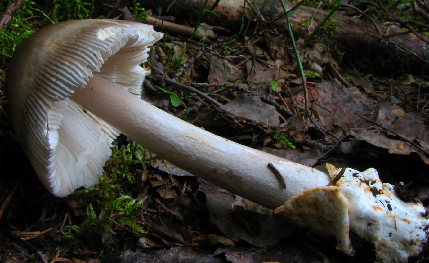
Irene Andersson (irenea) · CC BY-SA 3.0
Zmniejszono do maksymalnie 1600 pikseli i zapisano jako JPEG; zachowano proporcje, pełny kadr i barwy. Oryginał zachowano.
Photo source · CC BY-SA 3.0Amanita battarrae
Key differences
The grey amanita (Amanita vaginata) differs from the olive amanita by a cap of uniform shade without concentric zonation and a smooth, white stipe lacking darker scales.
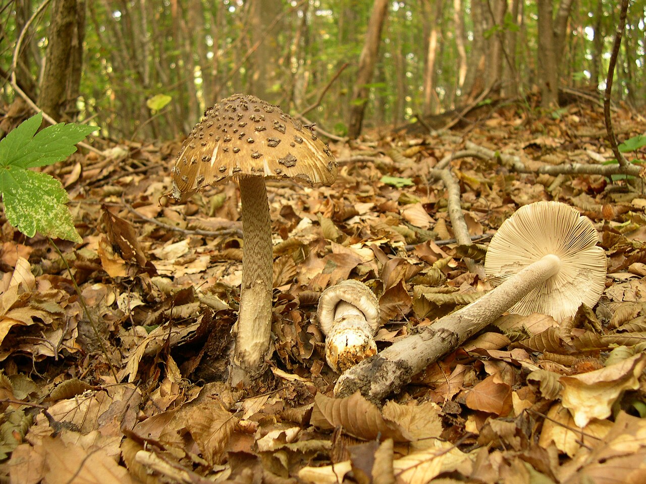
archenzo · CC BY-SA 3.0
Wersja JPEG o szerokości1280px przygotowana przez Wikimedia Commons; bez lokalnej zmiany kadru i koloru.
Photo source · CC BY-SA 3.0Amanita ceciliae
Key differences
The grey amanita (Amanita vaginata) is characterized by a smooth, white stipe devoid of dark scales and a zigzag pattern.
FAQ
Is Amanita vaginata edible?
Yes, it is edible when thoroughly cooked, but it is poisonous if consumed raw or undercooked.
What does the name 'grisette' mean?
The name derives from the French word for a grey woolen fabric worn by working-class women in 18th-century France.
Can this mushroom be eaten raw?
No, it contains heat-sensitive toxins that cause gastrointestinal illness unless neutralized by prolonged heat.
In which habitats does Amanita vaginata grow?
It occurs in deciduous, coniferous, and mixed woodlands, frequently near oaks, beeches, birches, and pines.
When is the fruiting season of Amanita vaginata?
Fruiting typically spans from mid-summer to late autumn, usually between June and November.
Does Amanita vaginata have a ring on its stem?
No, it lacks a partial veil and stem ring, which is a key defining characteristic of this species group.
Is this species used for dyeing textiles?
Artisanal textile crafters have used it with mordants to produce soft beige, grey, and olive tones on wool.
How is Amanita vaginata viewed in European culinary culture?
It is historically appreciated in French and Italian regional cuisine, but neglected in British culture.
Did 19th-century museums use models of this species?
Yes, wax and papier-mâché models were frequently cast for botanical education in colleges and museums.
What type of soil does Amanita vaginata prefer?
It tolerates acidic to neutral forest soils, ranging from sandy ground to rich loam covered in leaf litter.
Enthusiast
Names and history
- Obowiązująca nazwa krajowa
- muchomor szarawy
- Obowiązująca nazwa taksonomiczna
- Amanita vaginata (Bull.) Lam.
- Inne nazwy krajowe
- muchorówka bura, czubki, kania, kołpaczek, kurpytka popielata, cienkonóżka, pohnojka leśna, pniakówka pszeniczna, surojeżka siwa, bedłka mglejarka, maglejan, mglejara, bedłka pochwa, igielnica, Grisette
- Dawne nazwy krajowe
- bedłka pochwowata, bedłka, muchar pochwiasty, mglejarka pochwiasta, podsadka pochwiasta
- Pozostałe nazwy
- panienka, muchomor mglejarka
Home observations
Spore print
- Required materials
- fresh, mature cap, sheet of paper, glass or small bowl, water
- Time
- 2–24 h
- Procedure
- Place the cap underside down on the paper.
- Add a drop of water on top and cover.
- After 2–24 hours, lift the cap and inspect the deposit.
What to look for
- Spore print colour
- Spore print colour: white.
Under the microscope
- Required materials
- light microscope, microscope slide and cover slip, needle, water, spores from print
- Procedure
- Transfer a small amount of the spore print onto the slide using a needle.
- Add a drop of water, cover with a cover slip, and examine the spores.
What to look for
- Spore shape
- Spore shape: globose. Spore shape: subglobose. Spore shape: globose. Spore shape: globose. Spore shape: globose (nearly). Spore shape: globose (nearly).
- Spore surface
- Spore surface: smooth. Spore surface: smooth.
- Spore dimensions
- Length 9.2–13 µm, width 7.7–13 µm.
Compare dimensions after calibrating the microscope scale.
Ecology and distribution
- Soil reaction
- Soil reaction: acidic.
- Soil texture
- Soil texture: clayey.
Soil texture: loamy.
Soil texture: sandy. - Soil calcareousness
- Soil calcareousness: calcareous.
- Soil fertility
- Soil fertility: fertile.
- Broadleaf trees in the vicinity
- Broadleaf trees in the vicinity: beech.
Broadleaf trees in the vicinity: oak.
Broadleaf trees in the vicinity: birch.
Broadleaf trees in the vicinity: Sorbus aucuparia.
Broadleaf trees in the vicinity: poplars, including aspen. - Coniferous trees in the vicinity
- Coniferous trees in the vicinity: spruce.
Coniferous trees in the vicinity: Picea abies. - Shrubs in the vicinity
- Shrubs in the vicinity: Pinus mugo.
Shrubs in the vicinity: hazel. - Mycorrhizal hosts
- seedling inoculation experiment, field fruiting unconfirmed: Mycorrhizal hosts: Pinus tabuliformis (controlled experiment).
General species association: Mycorrhizal hosts: Betula (general partner list).
General species association: Mycorrhizal hosts: Corylus avellana (general partner list).
General species association: Mycorrhizal hosts: Populus tremula (general partner list).
General species association: Mycorrhizal hosts: Fagaceae (general partner list).
General species association: Mycorrhizal hosts: Pinaceae (general partner list). - Mode of nutrition
- Mode of nutrition: ectomycorrhizal.
Non-culinary uses
Documented non-culinary applications of Amanita vaginata are limited across ethnomycological records, yet several specific traditional and practical uses exist. In traditional fiber arts and textile dyeing experiments, dried or fresh caps have been utilized by artisanal crafters to yield subtle earth-toned natural dyes. When applied with alum or iron mordants on animal fibers such as wool and silk, extracts from the pigmented cap cuticle produce pale beige, muted tan, and dull greyish-olive shades, valued among fiber craft historians and historical reenactors.
Beyond natural dye production, the species possesses a history of utility within educational illustration, scientific modeling, and botanical display. Because of its distinctive anatomical features—such as the free gills, striated margins, and saccate basal volva—wax and papier-mâché models of Amanita vaginata were widely produced during the nineteenth and early twentieth centuries as standard physical teaching specimens for natural history museums and agricultural colleges. Additionally, in certain rural European folklore practices, the hollow stipes were occasionally hollowed further by children to fashion ephemeral woodland whistles.
History and culture
The grisette holds a distinctive place in classical European mycological history, having been formally described by French botanist Jean-Baptiste Bulliard in the late eighteenth century. Bulliard’s work established the species as a foundational taxon in early taxonomic literature. The common English name 'grisette' derives from the French word for an inexpensive grey woolen cloth, which in eighteenth- and nineteenth-century Parisian culture became associated with working-class young women; the mushroom’s muted grey coloration directly inspired the linguistic connection.
In regional folklore and rural agrarian traditions across southern and western Europe, the ringless amanitas acquired colorful folk names often referencing their sheathed, sock-like volval base or delicate umbrella form. In botanical art and Victorian natural history literature, Amanita vaginata was frequently rendered in hand-colored lithographs as an exemplar of woodland elegance and geometric balance. Cultural attitudes toward the species have historically mirrored deep European mycophilic and mycophobic divisions, with French and Italian rural folklore celebrating its seasonal appearance while Anglo-Saxon traditions viewed it with pervasive suspicion.
Edibility and safety
Amanita vaginata is an edible mushroom, but it is toxic when raw or insufficiently cooked, as it causes gastrointestinal distress and hemolysin-induced toxicity. Thorough heat processing is mandatory before consumption; prolonged boiling, thorough frying, or simmering ensures the breakdown of thermo-labile toxins. Consuming undercooked specimens carries substantial digestive risks, ranging from stomach cramps and nausea to vomiting. The flesh is fragile, tender, and mild-tasting, offering a delicate, somewhat sweet or nutty flavor profile once cooked.
Consumer appreciation varies considerably by region and culinary tradition. In France and Italy, the grisette is historically esteemed and regularly consumed in local communities where ringless amanitas have long held a place in rural diets. In parts of Central and Eastern Europe, it is also harvested for the table, though regarded by many as secondary due to its brittle flesh. In North America and Great Britain, culinary utilization is uncommon and frequently discouraged due to safety concerns regarding the genus, whereas in certain rural Asian localities, thoroughly cooked specimens are gathered as seasonal wild table mushrooms.
Habitat and occurrence
Amanita vaginata has a broad distribution across temperate and boreal zones of the Northern Hemisphere, occurring widely throughout Europe, North America, and temperate regions of Asia, with documented presence extending into subalpine zones. The fungus inhabits both broadleaf and coniferous woodlands, as well as mixed forests and heathland borders. It forms mycorrhizal associations with diverse tree species, notably oaks, beeches, birches, pines, spruces, and firs. It thrives in various soil types, tolerating acidic to neutral conditions, from loamy forest floors to sandy soils covered in leaf litter or moss cushions.
The fruiting season begins in mid-summer and continues into late autumn, typically spanning from June or July through October or November depending on latitude and altitude. In Mediterranean regions, fruiting often peaks later in autumn and can persist into early winter during mild, wet weather. In northern boreal and alpine regions, fruiting is compressed into a shorter window, peaking from late July through September. Fruiting is closely aligned with sustained soil moisture following summer rainfall.
Sources
- Commanster: Amanita vaginata
- Naturalis Interactive Guide: Amanita vaginata
- Karel Tejkal, Česká mykologická společnost: A.vaginata
- GRej, Amanita vaginata
- Stadt Wien, Marktamt: Grauer Scheidenstreifling
- Chu i in. 2021, Inoculation With Ectomycorrhizal Fungi…, DOI 10.3389/fmicb.2021.687304
- Wojewoda 2003, Checklist of Polish larger Basidiomycetes, s. 51
Researcher
Edibility and culinary use
Ustalenia źródłowe: jadalność, spożycie i wykorzystanie
- Edibility
- Edible — źródło oznaczenia
Zarzyński, Grzyby polskich lasów, s. 306–307 · p. 306 (PDF 304), Usability properties: edible mushroom; a prohibition against eating raw was explicitly noted.
- Raw consumption
- Poisonous
Materiał: surowy
Zarzyński, Grzyby polskich lasów, s. 306–307 · p. 306 (PDF 304), Usability properties: slight toxic properties before thermal processing.
- Required processing
- Requires preparation
Zarzyński, Grzyby polskich lasów, s. 306–307 · p. 306 (PDF 304), Usability properties; distinct contrast between raw and processed, without specifying minutes or temperature.
- Culinary rating
- Neutral
Zarzyński, Grzyby polskich lasów, s. 306–307 · p. 306 (PDF 304), Usability properties: without distinct taste or significant flavor qualities; atlas assessment, not a toxicological test.
Scientific names and taxonomy
- Current scientific name
Amanita vaginata (Bull.) Lam.
Scientific names and taxonomy
The correct scientific name is Amanita vaginata (Bull.) Lam., published in 1783 in Encyclopédie Méthodique, Botanique (volume 1(1), p. 109). The basionym is Agaricus vaginatus Bull., and in earlier treatments the combination Amanitopsis vaginata (Bull.) Roze was also used.
Historical scope of names and species concepts
In systematics, the taxon is assigned to the family Amanitaceae (Amanitaceae), although in historical classification indices it was sometimes placed in Agaricaceae (Agaricaceae).
Taxon scope and identification limitations
In the traditional, broad sense, the name Amanita vaginata encompassed a group of difficult-to-distinguish forms from section Vaginatae. Modern phylogenetic revisions based on molecular marker analysis (ITS, LSU, RPB1, TEF1-α) demonstrated the presence of several dozen distinct evolutionary lineages in this group and led to the delimitation of new, gray species such as A. deflexa, A. griseofulva, and A. opaca.
- Zakres ujęcia taksonomicznego
Amanita vaginata nomenclatural name IF114685; source-specific traditional broad atlas concepts, without assigning them to a molecularly delimited European A.vaginata sensu stricto. Indian ethnographic material retains its own broad attribution; Varga2024 section-level methods/new species are not results of this species.
[32] [1] [31]
Ecology and distribution
Laboratory diagnostics
Dane ustrukturyzowane
Basidia
- Clamp connections
- Clamp connections: absent. [55]
Hymenial cystidia
- Presence of cystidia in the hymenium
- Presence of cystidia in the hymenium: present. [51]
- Abundance of cystidia at the specified location
- Abundance of cystidia at the specified location: scattered. [51]
- Hymenial cystidium shape
- Hymenial cystidium shape: globose. [51]
Hymenial cystidium shape: pear-shaped. [51] - Hymenial cystidium size
- Hymenial cystidium size: large. [51]
Pileipellis
- Optical appearance of pileipellis hyphae
- Optical appearance of pileipellis hyphae: hyaline (in the lower pileipellis layer). [51]
- Arrangement of pileipellis hyphae
- Arrangement of pileipellis hyphae: Parallel (in the lower pileipellis layer, more or less). [51]
- Embedding of cuticle hyphae in a gelatinous substance
- Embedding of cuticle hyphae in a gelatinous substance: present (upper layer of the cap cuticle). [51]
Reactions with reagents
- Amyloid reaction
- Amyloid reaction: negative. [51]
Spores
Material and research methods
Description of characteristics — source and scope
Material: Assessment of flesh characteristics in university sheet Amanita vaginata; no specimen number or sensory panel.
Reading scope: Full text.
Limitations: Field guide assessment of odor, not a measurement of a chemical compound or an indication to taste the raw mushroom.
Microscopic data — source and limitations
Material: Aggregate spore result for A. vaginata var. vaginata in §3.2.10, Chinese material.
Reading scope: Full text.
Limitations: Qm is the reported mean of quotients, SD is not a min–max range. No calculation of Q from independent extreme lengths and widths.
Study of composition and activity — Malacara-Marquez et al. (2026)
Material: Methanolic extract of fruit bodies identified by the authors as A. vaginata, in a study covering six species. Only the qualitative result reported for A. vaginata and the MCF7 line was assigned.
Reading scope: Abstract.
Limitations: The abstract does not report species-specific IC50. The LD50 range from the entire panel or values of other species were not assigned. The in vitro result does not confirm human treatment or whole-meal toxicity.
Study of composition and activity — material and limitations
Material: EfraAva from dried fruit bodies identified as A. vaginata, Midnapur, West Bengal, India; voucher AMFH-507. Ethanolic extract; original proportions and conditions remain in the source method.
Reading scope: Full text.
Limitations: Morphological identification according to Pradhan et al. 2012; no DNA agreement with European material was demonstrated. In vitro extract study, without evidence of therapeutic effect or safety of a whole meal.
Study of composition and activity — Seliman et al. (2025)
Material: The same source purified lectin AVL, tested on LPS-stimulated murine macrophages RAW264.7. Protein expression levels were determined by Western blot, rather than direct enzyme activity.
Reading scope: Abstract.
Material application experiment — Yıldırım (2023)
Material: AVA: dry residue of fruit bodies after methanol extraction. AVAK: composite of this residue with sodium tripolyphosphate according to the authors. Material from Mardin, Turkey, without a specified voucher or DNA. Methylene blue removal was studied; stirring 120 rpm, kinetics study window 0–240 min. This is non-living material, not a fungal culture.
Reading scope: Full text.
Limitations: The authors reported experimental capacities and Qe,c from the fitted pseudo-second-order model separately. They were not combined into a range. The experiment indicates higher sorption of AVAK; the table of fitted Qe,c gives the opposite relationship. Both measures were preserved, without correcting the author's numbers. R² for AVAK at 150 mg/l differs between the table and text. Lack of evidence of industrial implementation.
Study of composition and activity — Drewnowska (2014)
Material: Caps, stems, and soil beneath fruit bodies from seven sites in Poland, harvested 2000–2008. Determination of mercury by CV-AAS at 253,7 nm, results on a dry weight basis. The source provides sample ranges and separate lower and upper site summaries.
Reading scope: Abstract.
Limitations: Available reading is an abstract; complete sample results and site names are lacking in this reading. Type of ± was not specified in the abstract. Sample boundaries were not derived from ± and dry weight was not converted to fresh weight. The BCF compares tissue with soil, and the cap/stem ratio compares two tissues. The results do not determine meal safety or remediation efficacy.
Study of composition and activity — Paloi and Acharya 2014 (2014)
Material: Ethanol fraction EfraAva from the same voucher AMFH-507. 30 g of powder was extracted with 500 ml of ethanol for 24 hours at 25°C, extraction was repeated, evaporated at 40°C. This preparation is not identical to AvaPre.
Reading scope: Full text.
Material preparation: EfraAva. 1. extraction — ethanol, 25 degrees Celsius, 24 hours. 2. re-extraction — ethanol, 25 degrees Celsius, 24 hours, repetition of step "ethanol-1". 3. drying, 40 degrees Celsius.
Limitations: Two articles use the same voucher, not two independent populations. Molecular conformity with the European type was not demonstrated. Equivalent values are results of specific chemical methods; TAC is not vitamin C content, and Folin does not identify gallic acid in the sample. The type of ± in the tabular composition summary was not added without explicit specification. Extract assays do not prove clinical effects or food safety.
Study of composition and activity — Paloi and Acharya 2013
Material: Multi-step polyphenolic fraction AvaPre from voucher AMFH-507, Midnapur, West Bengal, India. Morphological identification according to Pradhan et al. 2012. The structured procedure was preserved: ethanol, repeated extraction, dried filtrate, hot water, addition of ethanol, and preserved supernatant.
Reading scope: Full text.
Material preparation: AvaPre. 1. extraction — ethanol, 25 degrees Celsius, 2 days. 2. re-extraction — ethanol, 25 degrees Celsius, 2 days, repetition of step "ethanol-1". 3. drying; filtrate was retained. 4. extraction — water, 100 degrees Celsius, 8 hours. 5. addition — ethanol, 4 volumes relative to the "hot-water" step. 6. fraction separation; supernatant was retained.
Limitations: Two articles use the same voucher, not two independent populations. Molecular conformity with the European type was not demonstrated. Equivalent values are results of specific chemical methods; TAC is not vitamin C content, and Folin does not identify gallic acid in the sample. The type of ± in the tabular composition summary was not added without explicit specification. Extract assays do not prove clinical effects or food safety.
Plant inoculation experiment — material and limitations
Material: Experiment with Pinus tabulaeformis seedlings and inoculum of A. vaginata culture, obtained from a fruiting body collected in Huoditang, Qinling Mountains, China. Following colonization, plants infected with Bursaphelenchus xylophilus were compared with the corresponding control without fungal inoculation. The measured endpoint was rhizosphere microbial biomass carbon.
Reading scope: Full text.
Limitations: The text reports a relative difference of 38,16%. The description covers measurements 6 and 9 months after infection; no self-selected time point was assigned to this summary number. This is not a percentage of killed nematodes, proof of fruiting body cultivation, or a clinical outcome. Inoculation was not equated with an unconditional natural host relationship.
Molecular study — Seliman et al. (2025)
Material: Characterization of purified lectin AVL. The abstract describes 80% ammonium sulfate precipitation and affinity chromatography; the band mass was determined by SDS-PAGE. It was not considered a determination of quaternary structure.
Reading scope: Abstract.
Common limitations of source reading
Limitations: Reading limited to the abstract. Specific doses, specimen, and collection country were not provided in the available reading. Hemagglutination is not hemolysis. Efficacy of treatment in humans and the identity of AVL with the atlas hemolysin hypothesis were not demonstrated.
Archiwalny opis siedliska
Występuje w lasach pod bukami, dębami i świerkami, z którymi wchodzi w związki mikoryzowe.
[36]Archiwalny opis siedliska
Okres owocnikowania w Europie Środkowej trwa zasadniczo od maja do października, a pojedyncze znaleziska notowano jeszcze pod koniec listopada.
[28]- Results as recorded in the sources — Spores per basidium
- Liczba zarodników na jednej podstawce wynosi 4. [55] [51]
Metody i zapisy źródłowe — Własne udokumentowane obserwacje Josepa Torresa, Fungipedia. N=30 zarodników; nie podano numeru voucheru, daty ani jednoznacznego miejsca pobrania. Mapa GBIF nie jest geolokalizacją badanego okazu.
Metoda źródłowa. Technika: mikroskopia świetlna (LM). Opis metody w źródle: Odczytano opis i tabelę 30 pomiarów oraz etykiety galerii mikroskopowej. Nie dopisano jednego medium wszystkim cechom. Reakcję Melzera rozliczono osobno.
Wygląd optyczny strzępek skórki — zapis źródłowy: „Hialinas: wyłącznie zwarte strzępki pod suprapellis, bezbarwne optycznie; nie określono w ten sposób całej skórki.” [51]
Układ strzępek skórki — zapis źródłowy: „Strzępki pod suprapellis: dispuestas más o menos paralelamente.” [51]
Liczba zarodników na jednej podstawce — zapis źródłowy: „Basidios tetraspóricos.” [51]
Rozszerzenie podstawki — zapis źródłowy: „Basidios ... fuertemente ensanchados.” [51]
Kształt podstawki — zapis źródłowy: „Basidios claviformes; stopień rozszerzenia pozostaje odrębną nieobsługiwaną cechą.” [51]
Obecność cystyd w hymenium — zapis źródłowy: „Queilocistidios: cystydy wskazano konkretnie na ostrzu blaszki; nie wyciągnięto wniosku o ich obecności lub nieobecności na ścianach blaszki.” [51]
Obfitość cystyd — zapis źródłowy: „Queilocistidios dispersos.” [51]
Kształt cystydy hymenialnej — zapis źródłowy: „Queilocistidios ... entre globosos y piriformes.” [51]
Wielkość cystydy hymenialnej — zapis źródłowy: „Queilocistidios ... de gran tamaño; bez niepodanego wymiaru liczbowego.” [51]
Osadzenie strzępek skórki w substancji żelowej — zapis źródłowy: „Suprapellis filamentosa, formada por hifas finas, separadas por gelificación; obecność żelu w dokładnie nazwanej górnej warstwie, bez stopnia lub odsetka.” [51]
Krople wewnątrz zarodnika — zapis źródłowy: „Una gran gota en su interior; jedna duża kropla, bez wniosku o składzie lipidowym.” [51]
Długość zarodnika — zapis źródłowy: „Tabela esporas N=30, Largo: 9.21–12.98 µm; pełny zakres.” [51]
Iloraz długości i szerokości zarodnika — zapis źródłowy: „Tabela Q: 1.00–1.33; iloraz podany przez autora.” [51]
Kształt zarodnika — zapis źródłowy: „Esporas prácticamente esféricas; kuliste, z kwalifikatorem niemal przy tej wartości.” [51]
Powierzchnia zarodnika — zapis źródłowy: „Esporas de pared lisa.” [51]
Szerokość zarodnika — zapis źródłowy: „Tabela esporas N=30, Ancho: 7.70–11.83 µm; pełny zakres.” [51]
Metody i zapisy źródłowe — Materiał chiński z §3.2.10: sześć wymienionych kolekcji BJTC/HMAS. Zapis zarodników [60/3/3]; nie przypisano całego opisu pojedynczemu okazowi. Część oznaczeń ma sekwencje, dwa eksykaty bez uzyskanych sekwencji pozostają identyfikacją morfologiczną.
Metoda źródłowa. Technika: mikroskopia świetlna (LM). Opis metody w źródle: Suszony materiał, cienkie skrawki w 3% KOH lub wodzie, następnie 1% czerwień Kongo. Mikroskop Olympus DP71. Dla konkretnego zakresu nie wskazano jednego wyłącznego medium; nie dopisano go.
Liczba zarodników na jednej podstawce — zapis źródłowy: „§3.2.10: four-spored; pojedyncza liczba, bez przedziału alternatyw.” [55]
Długość podstawki; Szerokość podstawki — zapis źródłowy: „§3.2.10: basidia 48–60 × 13–18 µm.” [55]
Kształt podstawki — zapis źródłowy: „§3.2.10: clavate.” [55]
Długość zarodnika — zapis źródłowy: „§3.2.10: (9.2–)9.5–11.2(–13.0) µm.” [55]
Iloraz długości i szerokości zarodnika — zapis źródłowy: „§3.2.10: Q = (1.0–)1.02–1.08(–1.10).” [55]
Kształt zarodnika — zapis źródłowy: „§3.2.10: globose to subglobose; podstawowy kształt kulisty.” [55]
Kształt zarodnika — zapis źródłowy: „§3.2.10: subglobose; kulisty kształt z oddzielnym określeniem przybliżenia.” [55]
Szerokość zarodnika — zapis źródłowy: „§3.2.10: (8.0–)9.0–10.9(–13.0) µm.” [55]
Metody i zapisy źródłowe — GRej, Amanita vaginata
Metody i zapisy źródłowe — Karel Tejkal, Česká mykologická společnost: A.vaginata
Names and identification boundaries
Polish name: The primary name remains muchomor szarawy. Wojewoda lists earlier names: bedłka pochwowata, bedłka, muchar pochwiasty, mglejarka pochwiasta, and podsadka pochwiasta, indicating historical publications. Their presence attests to the use of the name in those studies, not a contemporary resolution of the identity of each historical specimen. Generic names, e.g. bedłka or kania from a folk atlas, may also refer to other fungi. They have been retained as alternative names with a source, without promoting them to the primary name.
Scientific concept: Amanita vaginata (Bull.) Lam. is a name based on Agaricus vaginatus Bull.; the former combination Amanitopsis vaginata remains in the history of names. In the analysis by Varga and co-authors from 2024, traditionally identified A. vaginata form several distant lineages. The authors defer a separate resolution of the typification problem and the European scope of this name. The traditional scope of the existing record has been retained; data from China and India are not presented as a molecularly confirmed Polish population sensu stricto.
Occurrence and season in Poland
Polish data: Wojewoda describes var. vaginata as common in Poland, growing on the ground in forests, thickets, parks, and mountain meadows. He mentions the vicinity of birches, Norway spruce, mountain pine, and rowan, without proof that every mentioned plant is a mycorrhizal partner. He reports June–October; the same VI–X range appears in the calendar of the State Forests' own field guide sheet. This is the Polish field guide season, distinct from individual collections and Indian dates.
Primary material: The mercury study by Drewnowska and co-authors included basidiocarps identified as A. vaginata from seven Polish sites, collected in 2000–2008. This is independent evidence of work on Polish material under this name. The abstract does not provide DNA determination of each specimen, so it does not resolve the modern boundaries of the complex. The historical frequency assessment from 2003 has not been equated with new national monitoring.
Microscopy — material from China
Material and method: Zhou and co-authors describe A. vaginata var. vaginata from the Yanshan Mountains. Six BJTC/HMAS collections are listed; the collective spore notation is [60/3/3]. Thin sections of dried material were observed in 3% KOH or water, then with 1% Congo red, using an Olympus DP71 microscope. The collective description is not attributed to a single specimen or a single exclusive medium. Part of the collections has sequences, two were based on morphology; the discrepancy in HMAS numbers 75237/75273 between sections of the article remains unresolved.
Results: The full outer limits of the spores are 9,2–13 × 8–13 µm, Q 1–1,10. The internal intervals of the source are retained as in the original. Qm 1,01 ± 0,03 is the mean with standard deviation, not an additional range. Spores are globose to subglobose. Basidia are 48–60 × 13–18 µm, clavate, and 4-spored. The authors state the absence of clamp connections in all examined tissues of the basidiocarps. The note that spore print observation was not conducted is not proof of the absence of a spore print.
Microscopy — Josep Torres's own observations
Material and scope: Torres provides a description and 30 spore measurements: 9,21–12,98 × 7,70–11,83 µm, Q 1–1,33. Lack of exsiccatum number, date, and unambiguous collection locality; the displayed GBIF map does not identify the examined specimen. No statistical significance was assigned to the unexplained abbreviation Q(Me). This is the author's primary observation, without DNA verification shown on this page. The Melzer's reaction result is negative; the absence of numerical reaction time and reagent dose is retained as a limitation.
Detailed structures: Spores are subglobose, smooth, with one large guttule; the author does not chemically identify it as a lipid. Cheilocystidia are scattered, large, globose to pyriform. Basidia are clavate, strongly widened, 4-spored. Hyphae of the suprapellis are separated by a gelatinous substance; the lower layer is more compact, with roughly parallel, hyaline hyphae without clamp connections. Layers, shape, size, degree, and approximation are retained separately. The observation of cheilocystidia does not establish the presence or absence of pleurocystidia.
Law in Poland — basis for the short result
Protection: The applicable official status and the full annexes of the regulation Dz.U. 2014 item 1408 were checked. Neither A. vaginata, the basionym, an alternative combination, nor a collective entry covering this species appears in the lists. Result: in Poland it is not subject to species protection. This is the result of a national act, distinct from the red list, protected area rules, and fungus identification.
Commercial trade: The consolidated text published on 3 March 2026, Dz.U. 2026 item 258, §2 section 1, and the entire Annex 1, items 1–72, were checked. The grisette and its proper scientific names do not appear on the list; it is not covered by a collective entry for the genus Amanita. Result: in Poland it is not permitted for commercial trade or the manufacture of mushroom products. This is not the same as a toxicity assessment.
Citation path and points where confirmation ends
Historical names: The examined Wojewoda list (2003, p. 51) refers for old Polish names to Kluk (1787), Jundziłł (1830), Chełchowski (1898), Błoński (1889, 1890), Kaufmann (1889), and Berdau (1876). The presence of these references in the list was confirmed; the indicated historical publications themselves were not read. Folk names in the State Forests atlas were read on p. 306, without attributing unstated primary informants to it. This history of names is not counted as a series of independent biological studies.
Identification of chemical material: Paloi and Acharya (2014), material and reference 20 → Pradhan and co-authors (2012), Amanita from Lateritic Region of West Bengal, Indian Forester 138(4):361–367, DOI 10.36808/if/2012/v 138 and 4/4638. Pradhan's abstract was read, not its paywalled full text. This reference documents the declared basis of the morphological identification; it does not replace reading the diagnosis or DNA sequences firsthand. Paloi 2014, reference 18 → Paloi and Acharya 2013: full text was read; both preparations retain the same voucher AMFH-507.
Claim of raw toxicity: Lyon card 1 → author's assessment regarding hemolysins and processing → no indicated reference to a specific primary experiment for this claim. State Forests Atlas → author's assessment of slight toxicity of raw fruiting bodies, without an experiment of their own. In these examined pathways, an identified hemolysin and a validated processing threshold were not established. The lack of a conclusive basis in the verified sequence does not prove the atlas assessment false nor the safety of the raw mushroom.
What constitutes an original experiment: Zhou 2023 → own material, microscopy, and sequences within the declared scope; Torres → own description and 30 measurements, without a stated voucher; Yıldırım 2023 → own biosorption experiment and parameter table; Chu 2021 → own plant inoculation experiment. The full corresponding descriptions were read. Result, method, material identification, and limitations are evaluated separately; primary source status alone does not eliminate table discrepancies or species identification limitations.
Limits of reviewing recent reports: Drewnowska and co-authors 2014 → available abstract of own Hg determination; Seliman and co-authors 2025/2026 → abstract of own AVL purification and cell experiment; Malacara-Marquez and co-authors 2026 → abstract of a six-species paper. Full protocols for these three papers were not read. Undisclosed doses, specimen numbers, specific IC50, or results for other species were not added. Data retain the level of an abstract, not full-text confirmation.
Occurrence verification — scope and limitations
Material: Polskie hasło Wojewody dla var. vaginata oraz pierwotny opis zbiorów z siedmiu polskich stanowisk z lat 2000–2008. Historyczna nazwa morfologiczna, bez potwierdzenia wszystkich okazów w nowoczesnym sensie molekularnym.
Scope of examination: Source excerpts.
A source excerpt was examined; full text and methods were not verified.
Review date: 18.10.2026.
Limitations: Identifications in historical sources concern their own interpretation of A. vaginata, rather than the epitypified European sensu stricto.
Occurrence verification — scope and limitations
Material: Chachuła 2018 wymienia gatunek w wynikach badań Dłubniańskiego Parku Krajobrazowego. Zachowano morfologiczne źródłowe ujęcie gatunku.
Reading scope: Source fragments.
A source fragment was read; full text and methods were not verified.
Review date: 21.10.2026.
Limitations: Source identification and conceptual scope retained. No new examination of specimens or reading of all publications cited by the source was declared.
Scope of elaboration
Required checks are settled.
Legal basis and scope of assessment
Analytical results
Composition and laboratory activity
- Activity of the tested extract
- Activity of the tested extract: antioxidant activity. [50]
Activity of the tested extract: cytotoxicity against cells (MCF7 cell line, extract, extract tested on the MCF7 cell line). [38]
Activity of the tested extract: antioxidant activity (extract, AvaPre extract). [38] - Quantitative content of component in material
- Quantitative content of component in material: 0.096 mg/kg (Hg, soil sample, unspecified measure of variation reported as ±, on a dry mass basis, soil for mercury determination, reported value after ±: 0.036, mean). [38]
Quantitative content of component in material: 0.028–0.47 mg/kg (Hg, fruit body preparation, on a dry mass basis, stipes for mercury determination, reported value). [38]
Quantitative content of component in material: 0.637 µg/mg (lycopene, extract, unspecified measure of variation reported as ±, on an extract mass basis, EfraAva extract, reported value after ±: 0.098, reported value). [38]
Quantitative content of component in material: 0.043–0.73 mg/kg (Hg, fruit body preparation, on a dry mass basis, caps for mercury determination, reported value). [38]
Quantitative content of component in material: 0.23 mg/kg (Hg, fruit body preparation, unspecified measure of variation reported as ±, on a dry mass basis, stipes for mercury determination, reported value after ±: 0.07, mean). [38]
Quantitative content of component in material: 1 µg/mg (β-carotene, extract, unspecified measure of variation reported as ±, on an extract mass basis, AvaPre extract, reported value after ±: 0.06, reported value). [38]
Quantitative content of component in material: 0.035 mg/kg (Hg, soil sample, unspecified measure of variation reported as ±, on a dry mass basis, soil for mercury determination, reported value after ±: 0.018, mean). [38]
Quantitative content of component in material: 0.48 mg/kg (Hg, fruit body preparation, unspecified measure of variation reported as ±, on a dry mass basis, caps for mercury determination, reported value after ±: 0.13, mean). [38]
Quantitative content of component in material: 0.729 µg/mg (ascorbic acid, extract, unspecified measure of variation reported as ±, on an extract mass basis, EfraAva extract, reported value after ±: 0.00031, reported value). [38]
Quantitative content of component in material: 0.017–0.16 mg/kg (Hg, soil sample, on a dry mass basis, soil for mercury determination, reported value). [38]
Quantitative content of component in material: 0.047 mg/kg (Hg, fruit body preparation, unspecified measure of variation reported as ±, on a dry mass basis, stipes for mercury determination, reported value after ±: 0.02, mean). [38]
Quantitative content of component in material: 0.6 µg/mg (lycopene, extract, unspecified measure of variation reported as ±, on an extract mass basis, AvaPre extract, reported value after ±: 0.03, reported value). [38]
Quantitative content of component in material: 1.7205 µg/mg (β-carotene, extract, unspecified measure of variation reported as ±, on an extract mass basis, EfraAva extract, reported value after ±: 0.265, reported value). [38]
Quantitative content of component in material: 0.00035 µg/mg (ascorbic acid, extract, unspecified measure of variation reported as ±, on an extract mass basis, AvaPre extract, reported value after ±: 0.00005, reported value). [38]
Quantitative content of component in material: 0.096 mg/kg (Hg, fruit body preparation, unspecified measure of variation reported as ±, on a dry mass basis, caps for mercury determination, reported value after ±: 0.052, mean). [38] - EC50 concentration in the specified antioxidant assay
- EC50 concentration in the specified antioxidant assay: 0.73 mg/ml (EC50, iron(II) ion chelation assay, extract, EfraAva extract, reported value). [38]
EC50 concentration in the specified antioxidant assay: 1.48 mg/ml (EC50, DPPH assay, extract, EfraAva extract, reported value). [38]
EC50 concentration in the specified antioxidant assay: 1.45 mg/ml (EC50, DPPH assay, extract, AvaPre extract, reported value). [38]
EC50 concentration in the specified antioxidant assay: 0.01 mg/ml (EC50, hydroxyl radical scavenging assay, extract, AvaPre extract, reported value). [38]
EC50 concentration in the specified antioxidant assay: 1.2 mg/ml (EC50, superoxide radical scavenging assay, extract, AvaPre extract, reported value). [38]
EC50 concentration in the specified antioxidant assay: 1.75 mg/ml (EC50, iron(II) ion chelation assay, extract, AvaPre extract, reported value). [38] - Reducing capacity in the Folin–Ciocalteu assay
- Reducing capacity in the Folin–Ciocalteu assay: 5.335 µgGAE/mg (Folin–Ciocalteu assay, extract, per mass of extract, unspecified measure of variation reported as ±, on an extract mass basis, AvaPre extract, calibration standard: gallic acid, reported value after ±: 0.28, reported value). [38]
Reducing capacity in the Folin–Ciocalteu assay: 11.296 µgGAE/mg (Folin–Ciocalteu assay, extract, per mass of extract, unspecified measure of variation reported as ±, on an extract mass basis, EfraAva extract, calibration standard: gallic acid, reported value after ±: 0.74, reported value). [38] - Mass-related antioxidant activity
- Mass-related antioxidant activity: 213 µgAAeq/mg (phosphomolybdenum assay, extract, per mass of extract, unspecified measure of variation reported as ±, on an extract mass basis, EfraAva extract, ascorbic acid, reported value after ±: 9.5, reported value). [38]
Mass-related antioxidant activity: 225 µgAAeq/mg (phosphomolybdenum assay, extract, per mass of extract, unspecified measure of variation reported as ±, on an extract mass basis, AvaPre extract, ascorbic acid, reported value after ±: 3.5, reported value). [38] - Flavonoid content in collective assay
- Flavonoid content in collective assay: 2.29 µgQE/mg (aluminium nitrate assay, extract, per mass of extract, unspecified measure of variation reported as ±, on an extract mass basis, EfraAva extract, quercetin, reported value after ±: 0.44, reported value). [38]
Flavonoid content in collective assay: 0.81 µgQE/mg (aluminium nitrate assay, extract, per mass of extract, unspecified measure of variation reported as ±, on an extract mass basis, AvaPre extract, quercetin, reported value after ±: 0.06, reported value). [38]
Biotechnological experiments
Activity of isolated compounds
- Activity of isolated compound
- Activity of isolated compound: reduced protein expression (RAW264.7 cell line, purified compound, dose-dependent, iNOS expression, AVL preparation tested on cells). [38]
Activity of isolated compound: reduced protein expression (RAW264.7 cell line, purified compound, dose-dependent, COX-2 expression, AVL preparation tested on cells). [38]
Sorption parameters
- Common study parameters
- Study conditions include temperature: 298 kelvins, sorbent mass: 0,01 grams, solution volume: 20 milliliters. [37]
- Model-fitted sorption capacity
- Model-fitted sorption capacity: 185.19 mg/g (methylene blue, pseudo-second-order model, calculated equilibrium sorption capacity Qe,c, composite, AVAK composite, reported value). [38]
Model-fitted sorption capacity: 769.23 mg/g (methylene blue, pseudo-second-order model, calculated equilibrium sorption capacity Qe,c, biomass residue, AVA biomass residue, reported value). [38]
Model-fitted sorption capacity: 90.09 mg/g (methylene blue, pseudo-second-order model, calculated equilibrium sorption capacity Qe,c, composite, AVAK composite, reported value). [38]
Model-fitted sorption capacity: 588.24 mg/g (methylene blue, pseudo-second-order model, calculated equilibrium sorption capacity Qe,c, biomass residue, AVA biomass residue, reported value). [38]
Model-fitted sorption capacity: 163.93 mg/g (methylene blue, pseudo-second-order model, calculated equilibrium sorption capacity Qe,c, composite, AVAK composite, reported value). [38]
Model-fitted sorption capacity: 714.29 mg/g (methylene blue, pseudo-second-order model, calculated equilibrium sorption capacity Qe,c, biomass residue, AVA biomass residue, reported value). [38] - Measured sorption capacity
- Measured sorption capacity: 76.6 mg/g (methylene blue, biomass residue, AVA biomass residue, reported value). [38]
Measured sorption capacity: 151.1 mg/g (methylene blue, composite, AVAK composite, reported value). [38]
Measured sorption capacity: 43.12 mg/g (methylene blue, biomass residue, AVA biomass residue, reported value). [38]
Measured sorption capacity: 133.58 mg/g (methylene blue, composite, AVAK composite, reported value). [38]
Measured sorption capacity: 129.76 mg/g (methylene blue, biomass residue, AVA biomass residue, reported value). [38]
Measured sorption capacity: 55.54 mg/g (methylene blue, composite, AVAK composite, reported value). [38] - Sorption model fit coefficient R²
- Sorption model fit coefficient R²: 0.9954 (methylene blue, pseudo-second-order model, calculated equilibrium sorption capacity Qe,c, biomass residue, source table, refers to model fit amanita_vaginata.w10.nr23.extensions.biosorption_fitted_capacity.1.result, AVA biomass residue, reported value). [38]
Sorption model fit coefficient R²: 0.9907 (methylene blue, pseudo-second-order model, calculated equilibrium sorption capacity Qe,c, biomass residue, source table, refers to model fit amanita_vaginata.w10.nr23.extensions.biosorption_fitted_capacity.2.result, AVA biomass residue, reported value). [38]
Sorption model fit coefficient R²: 0.09951 (methylene blue, pseudo-second-order model, calculated equilibrium sorption capacity Qe,c, composite, main text of the publication, refers to model fit amanita_vaginata.w10.nr23.extensions.biosorption_fitted_capacity.6.result, AVAK composite, reported value). [38]
Sorption model fit coefficient R²: 0.9859 (methylene blue, pseudo-second-order model, calculated equilibrium sorption capacity Qe,c, biomass residue, source table, refers to model fit amanita_vaginata.w10.nr23.extensions.biosorption_fitted_capacity.3.result, AVA biomass residue, reported value). [38]
Sorption model fit coefficient R²: 0.9951 (methylene blue, pseudo-second-order model, calculated equilibrium sorption capacity Qe,c, composite, source table, refers to model fit amanita_vaginata.w10.nr23.extensions.biosorption_fitted_capacity.6.result, AVAK composite, reported value). [38]
Sorption model fit coefficient R²: 0.9902 (methylene blue, pseudo-second-order model, calculated equilibrium sorption capacity Qe,c, composite, source table, refers to model fit amanita_vaginata.w10.nr23.extensions.biosorption_fitted_capacity.5.result, AVAK composite, reported value). [38]
Sorption model fit coefficient R²: 0.9898 (methylene blue, pseudo-second-order model, calculated equilibrium sorption capacity Qe,c, composite, source table, refers to model fit amanita_vaginata.w10.nr23.extensions.biosorption_fitted_capacity.4.result, AVAK composite, reported value). [38]
Element distribution
- Common study parameters
- Study conditions include the period from 2000 to 2008; number of sites: 7. [41]
- Element bioconcentration factor
- Element bioconcentration factor: 11 (Hg, ratio of mercury concentration in the cap to that in soil, unspecified measure of variation reported as ±, on a dry mass basis, upper value in the site summary, reported value after ±: 5, reported value). [38]
Element bioconcentration factor: 7.4 (Hg, ratio of mercury concentration in the stipe to that in soil, unspecified measure of variation reported as ±, on a dry mass basis, upper value in the site summary, reported value after ±: 3.9, reported value). [38]
Element bioconcentration factor: 0.61 (Hg, ratio of mercury concentration in the stipe to that in soil, unspecified measure of variation reported as ±, on a dry mass basis, lower value in the site summary, reported value after ±: 0.26, reported value). [38]
Element bioconcentration factor: 1.2 (Hg, ratio of mercury concentration in the cap to that in soil, unspecified measure of variation reported as ±, on a dry mass basis, lower value in the site summary, reported value after ±: 0.6, reported value). [38] - Ratio of element concentrations in cap and stem
- Ratio of element concentrations in cap and stem: 1.2–2.2 (Hg, ratio of mercury concentration in the cap to that in the stipe, on a dry mass basis, range of site medians). [38]
Ratio of element concentrations in cap and stem: 2.1 (Hg, ratio of mercury concentration in the cap to that in the stipe, unspecified measure of variation reported as ±, on a dry mass basis, upper value in the site summary, reported value after ±: 0.5, mean). [38]
Ratio of element concentrations in cap and stem: 1.2 (Hg, ratio of mercury concentration in the cap to that in the stipe, unspecified measure of variation reported as ±, on a dry mass basis, lower value in the site summary, reported value after ±: 0.4, mean). [38]
Plant experiments
- Relative change in parameter after plant inoculation
- Relative change in parameter after plant inoculation: 38.16 % (rhizosphere microbial biomass carbon, Pinus tabuliformis, increase, reported value). [38]
Characterization of purified protein
- Study material
- Material used — AVL. [49]
- Molecular mass of the tested protein
- Molecular mass of the tested protein: 45 kDa (purified compound, AVL preparation for protein characterization, reported value). [38]
- Protein purification fold
- Protein purification fold: 16 (purified compound, AVL preparation for protein characterization, reported value). [38]
- Protein purification yield
- Protein purification yield: 20 % (purified compound, AVL preparation for protein characterization, reported value). [38]
- Specific hemagglutinating activity of the protein
- Specific hemagglutinating activity of the protein: 5120 HU/mg protein (purified compound, AVL preparation for protein characterization, reported value). [38]
Test results — ferricyanide reduction
Test results — ferricyanide reduction
AvaPre — polyphenolic fraction from India, 2013
Material and preparation: Paloi and Acharya used basidiocarps from West Bengal, voucher AMFH-507, morphologically identified according to Pradhan and co-authors. AvaPre was obtained stepwise: extraction with ethanol at 25°C for two days, repeated, drying of the filtrate, hot water at 100°C for eight hours, addition of four volumes of ethanol, and retaining the supernatant. This is a specific laboratory fraction, not a whole meal or a preparation identical to EfraAva from a subsequent study.
Composition and activity: Per mg of extract, the following were reported: phenols 5,335 ± 0,28 µg gallic acid equivalent, flavonoids 0,81 ± 0,06 µg quercetin equivalent, β-carotene 1 ± 0,06 µg, lycopene 0,6 ± 0,03 µg, and ascorbic acid 0,00035 ± 0,00005 µg. Flavonoids were determined with aluminum nitrate. TAC was 225 ± 3,5 µg ascorbate equivalent/mg. EC50: hydroxyl radical 0,01; superoxide 1,2; DPPH 1,45; Fe 2+ chelation 1,75 mg/ml. A concentration of 1,55 mg/ml yielded an absorbance of 0,5 in the reducing power assay, not 50% inhibition.
EfraAva — ethanolic extract from India, 2014
Material and method: The same research group reused voucher AMFH-507 from Midnapur. For EfraAva, 30 g of powder was extracted with 500 ml of ethanol for 24 hours at 25°C, repeating the extraction; the solvent was evaporated at 40°C. This procedure is distinct from AvaPre. The two articles concern the same identified material, so they do not increase the number of independent populations. Lack of confirmation of DNA identity with European A. vaginata.
Results and limitations: Phenols: 11,296 ± 0,74 µg GAE/mg; flavonoids: 2,29 ± 0,44 µg QE/mg; β-carotene: 1,7205 ± 0,265 µg/mg; lycopene: 0,637 ± 0,098; ascorbic acid: 0,729 ± 0,00031. TAC: 213 ± 9,5 µg ascorbate equivalent/mg. EC50 DPPH: 1,48 mg/ml, Fe 2+ chelation: 0,73 mg/ml; absorbance 0,5 at 0,91 mg/ml. Figure captions describe three experiments performed with three replicates each; this does not mean nine populations. Chemical tests of the extract do not prove therapeutic efficacy or food safety.
Purified AVL lectin — cellular study
This is not proof of direct inhibition of these enzymes. The abstract of PMID 40679843 was reviewed (online 2025, volume 2026), without full doses or specimen data. Hemagglutination is not hemolysis; human therapeutic efficacy has not been demonstrated.
Methanol extract and MCF7
Scope of available information: It does not report a species-specific numerical value of IC50. Neither the Artemia test ranges for six species nor the phenolic results of another species were attributed to the grisette. This is a cellular finding, without evidence of antitumor efficacy in patients; the full article was not reviewed.
Mercury — Polish environmental material
Material, method, and ranges: Drewnowska, Nnorom, and Falandysz studied basidiocarps and soil from seven Polish sites from 2000–2008 using the CV-AAS method at 253,7 nm. They were not converted to fresh weight without moisture content.
Ratios and significance: BCF compares tissue to soil, while the cap/stipe ratio compares two tissues, which is why these are different indicators. Individual minimum and maximum, an off-the-shelf remediation method, or a safety assessment of a specific meal were not derived from them. Review limited to the abstract.
Extraction residue — dye biosorption
FTIR was used for characterization before and after sorption. This is a laboratory experiment on non-living material, not an implemented treatment plant.
Experimental results support the conclusion of greater AVAK sorption; the fitted values from the table reverse this relationship and clearly deviate from the experiment. These two statistics were not equated. The discrepancy of R² 0,09951 in prose and 0,9951 in the table was left without silent correction.
Pine inoculation — mycorrhiza experiment
Experimental design: Chu and co-authors (2021) obtained a culture of A. vaginata from a basidiocarp from Huoditang in the Qinling Mountains, propagated mycelium on PDA/PDB, and inoculated Pinus tabulaeformis. The plant–fungi system was studied following infection with the pine wood nematode Bursaphelenchus xylophilus. This was not cultivation of edible basidiocarps or a study of direct nematode mortality.
Key substances and their effects
Key substances and their effects
In biochemical studies conducted on fruiting bodies traditionally identified as Amanita vaginata in India (specimen AMFH-507), the properties of ethanol extracts were analyzed.
Key substances and their effects
The extract was found to contain polyphenols (11,3 µg GAE/mg), flavonoids (2,29 µg QE/mg), β-carotene (1,72 µg/mg), lycopene (0,64 µg/mg), and ascorbic acid (0,73 µg/mg).
Key substances and their effects
In laboratory tests, these extracts demonstrated antioxidant activity, iron chelating ability (EC50 = 0,73 mg/ml), and DPPH free radical scavenging activity (EC50 = 1,48 mg/ml).
Key substances and their effects
A specific lectin (AVL) with a molecular mass of 45 kDa and high hemagglutinating activity (5120 HU/mg) was isolated from the fruiting bodies.
Key substances and their effects
In in vitro cellular assays on macrophages, this lectin inhibited the expression of the pro-inflammatory enzymes iNOS and COX-2.
Key substances and their effects
Microscopic examinations of veil tissues and spore measurements within section Vaginatae confirm the need for precise analyses of volva sections to distinguish related species.
Edibility and risk — type of evidence
Polish field guide assessment: The State Forests field guide considers the species edible after processing, advising against raw basidiocarps as slightly poisonous; this is the guide author's assessment, not the result of their own toxicological experiment. Lack of distinct taste and brittleness are given as culinary traits. The sentence about the easily peelable cuticle in later Remarks refers to the compared A. fulva, so it was not transferred to the grisette. Nor were the soil or trees listed for another species attributed to it.
Hemolysin hypothesis: The Lyon 1 university sheet attributes thermolabile hemolysins to the raw species and mentions a temperature of 70°C. The page does not lead to a primary experiment identifying such a toxin in this material and validating a universal safety threshold. In this review, no such specific paper or unambiguous case report allowing the reconstruction of the entire chain was found. This is an evidential limitation, not a determination of falsehood of the guide's assessment. Hemagglutination of the purified lectin AVL was not equated with hemolysis.
Poisoning following misidentification: A case report from 2025 concerns a fatal poisoning by A. vidua mistaken for A. vaginata. The authors describe HPLC of amatoxins as well as macroscopic and microscopic identification of A. vidua. The incident documents the danger of misidentification, rather than a proven death from properly identified A. vaginata. The abstract was reviewed, without full clinical documentation.
Consumption tradition — field study
Interviews in eastern India: In an ethnobotanical study from 2023, A. vaginata has voucher SS18, the names Sal Chhatu and Budhi Onthe, 42 informants, RFC 0,27, and 123 use reports. Cooking and preparation with tamarind, garlic, and rice are described. This is a documented culinary practice of the respondents, not a controlled safety study. The Indian July–October season does not replace Polish data. The separate entry A. vaginata var. alba (SKM26) was not merged with SS18.
Ecological studies
Local frequency in studies of the Orawa-Nowy Targ Basin
Assessment method: Komorowska, Karasiński, and Heinrich (2023) distinguish three classes of local frequency: rare — up to 5 localities, frequent — 6–20, common — more than 20. A locality denotes one ATPOL grid square with a side of 1 km. The assessment covers the study area of the Orawa-Nowy Targ Basin along with the attached areas of the Przełom Białki pod Krempachami Nature Reserve and Pasieczny Las.
Result for Amanita vaginata: The frequent (6) entry indicates a class comprising 6–20 localities and six occupied squares: DG 38-06, DG 38-26, DG 38-33, DG 38-38, EG 30-25, and EG 30-32. The number 6 does not specify the number of fruiting bodies or herbarium collections. Multiple collections from a single square are counted as one locality; the question mark next to EG 30-25 indicates an imprecise location.
Source documentation
Source findings: Naturalis describes Amanita vaginata in deciduous forests and avenues, especially with poplars, as well as oaks, birches, and hazel. It lists clayey and loamy soils as well as rich or calcareous sands. It gives the period summer–autumn. This qualitative season was retained for the scope of the Dutch description; it was not converted to the Polish season, and avenues were not equated with natural forest. The "Kwetsbaar" assessment is a red list status, distinct from species protection.
Habitat and occurrence
Potwierdzony zasięg występowania
Wszystkie obszary potwierdzone w zapisanych danych źródłowych.
| Kraj | Regiony i stanowiska | Fruiting | Sources |
|---|---|---|---|
| Poland | Dłubnia Landscape Park | VI–X |
|
Sources
- PHYLO-VAGINATAE-EUROPE-2024
- Komorowska H., Karasiński D., Heinrich Z. 2023 (author order on title page). The Agaricomycotina of the Kotlina Orawsko-Nowotarska basin, Western Carpathian Mts (Poland). W. Szafer Institute of Botany, Polish Academy of Sciences. ISBN 978-83-62975-46-4; DOI 10.35535/978-83-62975-46-4.
- Naturalis Interactive Guide: Introduction
- Naturalis Interactive Guide: Amanita vaginata
- Paloi i Acharya 2013, Int. J. PharmTech Res. 5(4): 1645–1654 — uporządkowane wyniki tego samego badania (także [46])
- Paloi i Acharya 2014, Asian J. Pharm. Clin. Res. 7(2): 88–92 — uporządkowane wyniki tego samego badania (także [47])
- Pierwotny zapis źródłowy — P0: amanita_vaginata
- Karel Tejkal, Česká mykologická společnost: A.vaginata
- Minister Środowiska, Dz.U. 2014 poz. 1408, rozporządzenie w sprawie ochrony gatunkowej grzybów
- Minister Zdrowia, Dz.U. 2026 poz. 258, tekst jednolity rozporządzenia dotyczącego grzybów dopuszczonych do obrotu
- GRej, Amanita vaginata
- Index Fungorum record114685
- Soumitra Paloi, Krishnendu Acharya 2014, Asian J.Pharm.Clin.Res.7(2): 88–92, Evaluation of antioxidative activity and chemical composition...
- Seliman T.M.H. i in., Natural Product Research40(15): 4557–4565(2026), epub 18.07.2025 DOI 10.1080/14786419.2025.2530212 (także [49])
- Stadt Wien, Marktamt: Grauer Scheidenstreifling
- Yıldırım, Polat, Aktay i Acay 2023, Kadirli 3(2): 285–298
- Chu i in. 2021, Inoculation With Ectomycorrhizal Fungi…, DOI 10.3389/fmicb.2021.687304
- Badanie ilościowe dzikiej żywności plemion wschodnich Indii, 2023, DOI 10.3389/fsufs.2023.1198187
- Drewnowska, Nnorom i Falandysz 2014, Mercury in the Grisette, DOI 10.1080/03601234.2014.896677
- Muchomor szarawy — aktualne polskie wykazy ochrony i obrotu
- Zarzyński, Grzyby polskich lasów, s. 306–307
- Université Lyon 1, Champignons: Amanita vaginata
- Malacara-Marquez i in. 2026, Bioactive metabolites from edible fungi, DOI 10.1080/10826068.2026.2640061
- ELI, obowiązujący status Dz.U. 2014 poz. 1408
- Muchomor szarawy — udokumentowane wyniki źródłowe
- Josep Torres, własne obserwacje i 30 pomiarów zarodników A. vaginata, Fungipedia
- ELI, obowiązujący status Dz.U. 2026 poz. 258
- Opis przypadku zatrucia A. vidua, błędnie uznaną za A. vaginata, PMID 40819824
- Wojewoda 2003, Checklist of Polish larger Basidiomycetes, s. 51
- Zhou i in. 2023, Diversity and taxonomy of Amanita in the Yanshan Mountains, DOI 10.3389/fpls.2023.1226794