
Edible
Requires preparation

Poisonous when raw

Set your location to see data for your region.


Amanita crocea, known as the saffron ringless amanita or orange grisette, is an edible agaric in the Amanita section Vaginatae. It is celebrated for its striking bright saffron-orange to apricot cap with furrowed striate margins, a tall, slender stem adorned with orange-buff zig-zag chevron bands, and a prominent, sheath-like white volva, growing in mycorrhizal association with various deciduous and coniferous forest trees across temperate regions.
Identification, habitat, and key differences.
Forager
Fruitbody structure
- Fruit body form
- Fruit body form: Cap, stipe.
Fruit body form: Cap.
Cap
- Cap shape
- Cap shape: hemispherical.
Cap shape: ovoid.
Cap shape: bell-shaped.
Cap shape: convex.
Cap shape: flat. - Cap surface relief
- when dry: Cap surface relief: smooth.
Cap surface relief: smooth. - Cap covering
- when dry: Cap covering: suede-like.
- Cap surface sheen
- Cap surface sheen: shiny.
- Cap stickiness and sliminess
- when wet: Cap stickiness and sliminess: sticky.
- Cap colour
- Cap colour: orange-ochre.
Cap colour: yellow-orange.
Cap colour: orange.
Cap colour: orange-brown.
Cap colour: orange-yellow. - Cap margin surface relief
- Cap margin surface relief: grooved.
- Cap diameter
- The cap measures 30–120 mm in diameter.
- Cap centre
- Cap centre: with a distinct umbo.
Cap underside
- Hymenophore type
- Hymenophore type: gills.
Gills at stipe
- Hymenophore attachment to the stipe
- Hymenophore attachment to the stipe: free from the stipe (gills).
- General gill profile
- General gill profile: ventricose (gills).
- Hymenophore colour
- Hymenophore colour: creamy white.
Hymenophore colour: saffron yellow.
in mature fruit bodies: Hymenophore colour: yellowish.
in young fruit bodies: Hymenophore colour: whitish.
Hymenophore colour: cream. - Macroscopic structure of the free gill edge
- in young fruit bodies: Macroscopic structure of the free gill edge: pruinose.
- Gill spacing
- Gill spacing: crowded.
- Gill thickness
- Gill thickness: thin.
- Gill width class
- Gill width class: broad.
- Presence of short gills
- Presence of short gills: present.
Stem and base
- Stipe position
- Stipe position: central.
- Stipe shape
- Stipe shape: cylindrical.
Stipe shape: tapering upwards. - Stipe colour
- Stipe colour: white.
Stipe colour: cream-yellow. - Stipe surface covering
- Stipe surface covering: floccose.
Stipe surface covering: scaly. - Stem surface pattern
- Stem surface pattern: zigzag.
- Colour of stipe ornamentation
- Colour of stipe ornamentation: orange.
Colour of stipe ornamentation: yellowish.
Colour of stipe ornamentation: yellow-orange. - Stipe interior
- Stipe interior: hollow.
in young fruit bodies: Stipe interior: solid. - Stipe consistency
- Stipe consistency: brittle.
- Stipe length
- Stipe length: 100–180 mm.
- Stipe diameter
- Stipe diameter: 12–30 mm.
in the lower part of the stipe: Stipe diameter: 20–25 mm.
in the upper part of the stipe: Stipe diameter: 10–15 mm. - Volva
- Volva: present.
Volva: present.
Volva: present.
Ring
- Ring presence
- Ring presence: no ring.
Universal veil at base
- Veil structure at the stipe base
- Veil structure at the stipe base: sac-shaped.
- Softness and firmness of the volva and veil at the base
- Softness and firmness of the volva and veil at the base: firm (veil structure at the stem base: volva).
- Material of volva and veil at stem base
- Material of volva and veil at stem base: membranous (veil structure at the stem base: volva).
Flesh
- Flesh colour
- Flesh colour: whitish.
Flesh colour: white.
Flesh colour: yellowish white.
Flesh colour: orange (cap flesh, beneath the surface layer).
Flesh colour: orange-brown (cap flesh, beneath the surface layer). - Flesh taste
- Flesh taste: weak.
- Flesh resistance to breaking or tearing
- Flesh resistance to breaking or tearing: brittle.
- Flesh odour
- Flesh odour: mushroom-like.
Flesh odour: pleasant (flesh).
Flesh odour: not distinctive (flesh).
Flesh odour: not distinctive (flesh). - Flesh flavour note
- Flesh flavour note: nutty.
- Distinctiveness of flesh taste
- Distinctiveness of flesh taste: indistinctive.
Distinctiveness of flesh taste: indistinctive. - Assessment of flesh taste pleasantness
- Assessment of flesh taste pleasantness: pleasant.
- Flesh thickness class
- Flesh thickness class: thin.
Where and when it grows
- Habitat
- Habitat: deciduous woodland.
Habitat: mixed woodland.
Habitat: park. - Substrate
- Substrate: soil.
- Fruit body growth pattern
- Fruit body growth pattern: solitary.
Fruit body growth pattern: in groups. - Occurrence frequency
- Occurrence frequency: rare.
- Fruiting season
- Fruit bodies appear from July to September.
- Distribution area
- Distribution area: confirmed.
Edibility and culinary use
- Edibility
- Edible
- Required processing
- Requires preparation
- Raw consumption
- Poisonous when raw
Law
- Protection
- It is not subject to species protection in Poland.
- Trade
- It is approved in Poland for neither trade nor the production of mushroom products.
Possible confusions
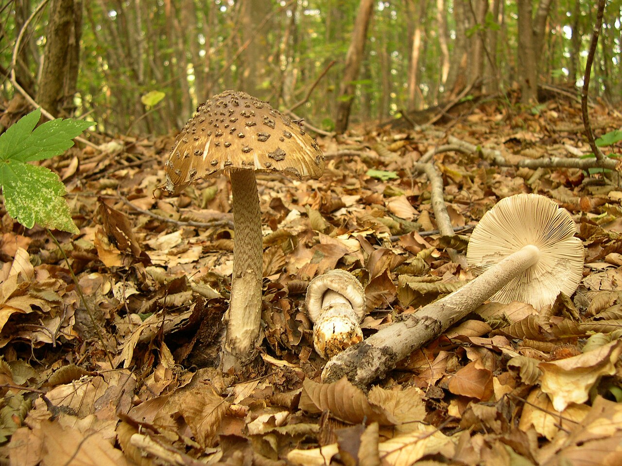
archenzo · CC BY-SA 3.0
Wersja JPEG o szerokości1280px przygotowana przez Wikimedia Commons; bez lokalnej zmiany kadru i koloru.
Photo source · CC BY-SA 3.0Amanita ceciliae
Key differences
The saffron ringless amanita (Amanita crocea) has a vivid saffron to orange-yellow cap and a volva colored orange on the inside.
FAQ
Is Amanita crocea safe to eat?
It is edible and prized once thoroughly cooked, but it is toxic and causes digestive distress if eaten raw.
What does 'crocea' mean in its scientific name?
The specific epithet comes from the Latin word 'croceus', meaning saffron-colored or golden-yellow.
Why must Amanita crocea be thoroughly heated?
It contains heat-labile toxins that disrupt red blood cells and digestion, which heat completely destroys.
In what forest types does Amanita crocea grow?
It occurs in deciduous, mixed, and coniferous woodlands, associating with birch, beech, oak, and pine.
When does Amanita crocea typically fruit?
Its fruiting season spans from early summer to late autumn, usually between July and November.
Does Amanita crocea have a ring on its stalk?
No, like other species in section Vaginatae, it lacks a partial veil and stem ring.
Can Amanita crocea be used as a textile dye?
Yes, textile artisans use cap pigments with mineral mordants to dye wool and silk golden-tan and yellow.
What soil conditions does Amanita crocea favor?
It favors acidic, nutrient-poor substrates, peat borders, heathlands, and mossy forest edges.
Where is this mushroom culturally popular as food?
It is traditionally esteemed and gathered in Italy, France, and parts of the Iberian Peninsula.
Was Amanita crocea used in historical museum displays?
Yes, wax and freeze-dried models were crafted for forestry and natural history exhibits to study fungi.
Enthusiast
Names and history
- Obowiązująca nazwa krajowa
- muchomor żółtawy
- Obowiązująca nazwa taksonomiczna
- Amanita crocea (Quél.) Singer
- Inne nazwy krajowe
- Orange Grisette
- Pozostałe nazwy
- muchomor żółty
Home observations
Spore print
- Required materials
- fresh, mature cap, sheet of paper, glass or small bowl, water
- Time
- 2–24 h
- Procedure
- Place the cap underside down on the paper.
- Add a drop of water on top and cover.
- After 2–24 hours, lift the cap and inspect the deposit.
What to look for
- Spore print colour
- Spore print colour: white.
Under the microscope
- Required materials
- light microscope, microscope slide and cover slip, needle, water, spores from print
- Procedure
- Transfer a small amount of the spore print onto the slide using a needle.
- Add a drop of water, cover with a cover slip, and examine the spores.
What to look for
- Spore shape
- Spore shape: globose. Spore shape: subglobose. Spore shape: globose. Spore shape: globose. Spore shape: globose. Spore shape: globose (nearly).
- Spore surface
- Spore surface: smooth. Spore surface: smooth. Spore surface: smooth.
- Spore dimensions
- Length 8.45–14.5 µm, width 7.42–11.2 µm.
Compare dimensions after calibrating the microscope scale.
Phenol experiment
- Material
- Stipe context
- Reagent
- phenol
- Procedure
- In the described trial, phenol was applied to the indicated material.
- Conditions and source gaps
The source does not provide: sample preparation, phenol concentration, reagent amount, temperature, reading time, detailed method of reagent application.
Observation / result
- Source result
- In the stem flesh, after application of phenol, a color described in the source as "brownish-wine" was obtained.
Ecology and distribution
- Soil reaction
- Soil reaction: acidic.
- Broadleaf trees in the vicinity
- Broadleaf trees in the vicinity: birch.
Broadleaf trees in the vicinity: oak. - Coniferous trees in the vicinity
- Coniferous trees in the vicinity: spruce.
- Mycorrhizal hosts
- General species association: Mycorrhizal hosts: Betula (general partner list).
General species association: Mycorrhizal hosts: Picea (general partner list). - Mode of nutrition
- Mode of nutrition: ectomycorrhizal.
Non-culinary uses
While non-culinary applications of Amanita crocea are rare in commercial industry, the species has specific documented historical and traditional uses in craft, natural pigment extraction, and education. Natural fiber artisans and botanical textile dyers have historically used the brightly pigmented pileipellis of fresh or dried caps to produce warm earth-toned dye baths. When combined with mineral mordants such as alum, copper, or iron salts, wool and unbleached silk fibers absorb soft golden-yellow, apricot-tan, and mustard hues, making the mushroom a historical dye source in traditional northern and alpine European textile craft.
In educational and scientific contexts, Amanita crocea has served a long-standing functional role in mycological pedagogy. Because of its vivid ornamentation—displaying bright cap pigmentation alongside distinct zig-zag stipe patterning and a prominent volva—it was frequently selected for creating botanical models, wax replicas, and taxidermic freeze-dried dioramas in university collections, forestry institutes, and natural history exhibits to teach fungal morphology and forest biodiversity without relying on perishable field material.
History and culture
Amanita crocea was recognized in early European mycology as a vibrant variety of the grisette group before receiving formal species recognition. The specific epithet 'crocea' derives from the Latin 'croceus', meaning saffron-colored or golden-orange, referring to the vivid coloration of saffron crocuses. This vibrant hue earned the mushroom a distinguished reputation in classical continental mycology, where eighteenth- and nineteenth-century naturalists singled it out as one of the most visually captivating forest fungi.
In European cultural heritage, particularly in rural French, Italian, and Swiss alpine valleys, the saffron ringless amanita is woven into regional folklore under colorful vernacular names such as 'couteau d'or' (golden knife) or 'angelot'. In historical botanical illustration, the elegant stature and warm glowing colors of the mushroom made it a favored subject for Victorian mycological plates and hand-painted instructional posters used in European village schools to teach local flora. Symbolically, its striking orange cap emerging through dark woodland moss was traditionally interpreted in alpine folklore as a herald of midsummer warmth yielding to autumnal harvest.
Edibility and safety
Amanita crocea is an edible species of good culinary quality, but like its close relatives, it contains heat-labile hemolytic toxins that render it poisonous in the raw state. Consuming raw or undercooked fruitbodies triggers acute gastrointestinal distress, including vomiting, abdominal pain, and cramping. Complete heat treatment is strictly required to neutralize these toxins; boiling, steaming, or comprehensive sauteing must be sustained until the flesh is cooked completely through.
Once properly cooked, the flesh offers a pleasant, delicate flavor with sweet, nutty undertones and a tender, brittle texture. Consumer acceptance varies noticeably across geographical borders. In Italy, France, and parts of the Iberian Peninsula, it is held in high gastronomic regard and sought after in local markets and village kitchens. In Central and Eastern European traditions, it is gathered and valued for its flavor, though handled with care due to its fragile structure. Conversely, British and North American culinary traditions largely overlook or discourage its consumption out of abundance of caution toward wild amanitas.
Habitat and occurrence
Amanita crocea is widely distributed throughout temperate, boreal, and subalpine regions of Europe and temperate Asia, with closely related taxa documented in North America. It displays a strong preference for mixed and deciduous woodlands, though it also establishes flourishing populations in coniferous stands. Common tree associates include birch, beech, oak, sweet chestnut, hazel, spruce, and pine. The species exhibits a pronounced affinity for acidic, nutrient-poor substrates, frequently fruiting among acidic humus, peat borders, heathlands, sphagnum edges, and sandy or gravelly forest paths.
Its fruiting season commences in early summer and extends through mid-to-late autumn, typically spanning from July to October or November depending on regional climate. In northern European birch forests and subalpine zones, sporocarps emerge predominantly during the warmer weeks of August and September. In warmer Mediterranean and southern European forests, fruiting peaks significantly later, often during October and November when autumn rains replenish damp woodland litter.
Researcher
Edibility and culinary use
Ustalenia źródłowe: jadalność, spożycie i wykorzystanie
- Required processing
- Requires preparation
Tejkal, Česká mykologická společnost, Amanita crocea · Jedlá, nutná delší tepelná úprava; author culinary assessment, not an experimentally determined safe procedure.
- Edibility
- Edible — źródło oznaczenia
Materiał: po obróbce
Tejkal, Česká mykologická společnost, Amanita crocea · CMS:jedlá, nutná delší tepelná úprava. Author assessment after cooking, not proof of safety of the raw mushroom.
- Raw consumption
- Poisonous
Materiał: surowy
Źródło bez opisu bibliograficznego · Remarque; Syndrome associé: harmful haemolytic effect in absence of cooking. Species page author assessment, not a validated preparation protocol.
Scientific names and taxonomy
- Current scientific name
Amanita crocea (Quél.) Singer
Scientific names and taxonomy
The saffron ringless amanita (Amanita crocea (Quél.) Singer) belongs to the family Amanitaceae (Amanitaceae). The basionym of the taxon is Amanita vaginata var. crocea Quél., described in 1898. The current combination at the species level was established by Rolf Singer in a work from 1951 (dated in sources to 1949–1951).
Taxon scope and identification limitations
Molecular studies of European representatives of section Vaginatae distinguish Amanita crocea in the narrow sense from Amanita subnudipes. Earlier collections from thermophilous oak forests on limestone, described as A. crocea, in reality correspond to A. subnudipes. True Amanita crocea sensu stricto occurs on acidic substrates under birches and spruces, characterized by a distinct orange hue of the stem.
Taxon scope and identification limitations
Modern taxonomic analyses of section Vaginatae are based on combined sequencing of the ITS, LSU, RPB1, and TEF1-α loci, as well as microscopic analysis of the anatomical structure of the volva.
Taxon scope and identification limitations
In measurements of fresh specimens from the Middle East, the mean mass of a fruit body was 73,74 ± 1,50 g, the stem length was 10,28 ± 0,67 cm, and the mean cap dimensions were approximately 7,5–9 cm.
- Zakres ujęcia taksonomicznego
Amanita crocea IF115109; source-specific atlas concepts and separate identified experimental materials. Varga2024 A.crocea sensu stricto remains distinct from historical Hungarian oak/calcareous records referred to A.subnudipes; Iranian isolate and Iraqi Dukan material retain only their own source identifications.
[26] [4]
Ecology and distribution
- Occurrence frequency
- Poland: Occurrence frequency: rare. [41]
Laboratory diagnostics
Dane ustrukturyzowane
Basidia
Hymenial cystidia
Pileipellis
Reactions with reagents
Spores
- Median of spore length to width ratio
- Median of spore length to width ratio: 1.2 (sample count: 30, median). [38]
- Spore colour under the microscope
- Spore colour under the microscope: hyaline. [46]
Spore colour under the microscope: hyaline. [50] - Spore length
- Spore length: 10–12 µm. [25]
Spore length: 8.45–11.24 µm. [38]
Spore length: 10.5–14.5 µm. [46] - Spore length-to-width ratio
- Spore length-to-width ratio: 1.03–1.37. [38]
- Spore shape
- Spore shape: globose. [25]
Spore shape: subglobose. [25]
Spore shape: globose. [46]
Spore shape: globose. [38]
Spore shape: globose. [50]
Spore shape: globose (nearly). [34] - Spore wall thickness
- Spore wall thickness: thin-walled. [38]
Material and research methods
Chemical reagent test — material and limitations
Material: Stem flesh of the fruit body in the SMNF account. No specific collection used for the test, reagent concentration, or reading time was provided.
Reading scope: Full text.
Limitations: Chocolate-brown color in the comparison applies to A. fulva; it was not assigned to A. crocea.
Microscopic data — source and limitations
Material: Josep Torres's own study in Fungipedia: 30 spores. No unequivocal collection number or geolocation of the studied specimen was provided.
Reading scope: Full text.
Limitations: Median values L 9,80 µm and W 8,52 µm were retained in the documentation; they do not replace the source min–max.
Composition and activity study — No. 24 — material
Material: Turkey: Beykonağı, Ilgın, Konya; pine forest, June 2018. Approximately 10 fruit bodies were collected; five were used for extraction after drying for 48 hours at 40°C. The remaining material was preserved in the fungarium of Selçuk University. Macro- and microscopic identification, without own DNA redetermination.
Reading scope: Full text.
Limitations: Three determination replicates are not three independent populations. A negative mutagenicity result and antimutagenicity in a bacterial assay do not confirm food safety or efficacy in humans. The source name "Hinic acid" was retained without arbitrary identification with quinic acid.
- Results as recorded in the sources — Spores per basidium
- Liczba zarodników na jednej podstawce wynosi 4. [46] [38] [50]
Metody i zapisy źródłowe — GarcíaBona, własny materiał z Nawarry, hiszpański opis skórki, osłony, ostrza, podstawek i zarodników. Zapis10.5[12.1;12.9]14.5×8.9[9.8;10.2]11.2µm zachowany, bez zgadywania statystyki liczb w nawiasach. Brak kalibracji, N i DNA.
Identyfikacja kropli: nierozstrzygnięta.
Układ strzępek skórki — zapis źródłowy: „Hifas entremezcladas; splątany układ strzępek.” [46]
Liczba zarodników na jednej podstawce — zapis źródłowy: „Basides tétrasporiques/basidios producen cuatro esporas;4na1podstawce.” [46]
Kształt podstawki — zapis źródłowy: „Podstawki wąskie u podstawy i rozszerzone ku górze; maczugowaty podstawowy kształt, bez zgadywania wymiarów.” [46]
Kształt cystydy hymenialnej — zapis źródłowy: „Queilocistidios claviformes o piriformes: maczugowata możliwość, tylko cystydy ostrza. Brakujące gruszkowate zapisano w dokumentacji.” [46]
Przegrody strzępki skórki — zapis źródłowy: „Tabiques desprovistos de fíbulas; jawnie opisane przegrody.” [46]
Osadzenie strzępek skórki w substancji żelowej — zapis źródłowy: „Strzępki skórki silnie osadzone/rozdzielone przez żel. Obecność oddzielona od nieobsługiwanego stopnia.” [46]
Barwa pigmentu skórki — zapis źródłowy: „Intracellular pigment: amarillento. Barwa i położenie osobno.” [46]
Barwa zarodnika w mikroskopie — zapis źródłowy: „Hyalines/incoloras; bezbarwne zarodniki w mikroskopii, nie barwa wysypu.” [46]
Zawartość zarodnika — zapis źródłowy: „Gran vacuola central; zawartość wakuolizowana. Nie zidentyfikowano lipidów i nie zapisano kropli oleju.” [46]
Długość zarodnika; Szerokość zarodnika — zapis źródłowy: „GarcíaBona, własny materiał z Nawarry, hiszpański opis skórki, osłony, ostrza, podstawek i zarodników. Zapis10.5[12.1;12.9]14.5×8.9[9.8;10.2]11.2µm zachowany, bez zgadywania statystyki liczb w nawiasach. Brak kalibracji, N i DNA.” [46]
Kształt zarodnika — zapis źródłowy: „Źródłowo globose/sphériques/téměřkulovité, podstawowy kulisty kształt.” [46]
Powierzchnia zarodnika — zapis źródłowy: „Esporas lisas; gładka powierzchnia zarodników, nie cystyd.” [46]
Metody i zapisy źródłowe — JosepTorres,Fungipedia,własny pomiar N=30zarodników. L8.45–11.24µm,W7.42–10.25µm,Q1.03–1.37; środkowe9.80,8.52 nie są skrajnymi granicami. MapaGBIF nie stanowi lokalizacji badanego okazu. Nie podano DNA.
Identyfikacja kropli: nierozstrzygnięta.
Liczba zarodników na jednej podstawce — zapis źródłowy: „Basides tétrasporiques/basidios producen cuatro esporas;4na1podstawce.” [38]
Kształt podstawki — zapis źródłowy: „Podstawki wąskie u podstawy i rozszerzone ku górze; maczugowaty podstawowy kształt, bez zgadywania wymiarów.” [38]
Kształt cystydy hymenialnej — zapis źródłowy: „Queilocistidios claviformes o piriformes: maczugowata możliwość, tylko cystydy ostrza. Brakujące gruszkowate zapisano w dokumentacji.” [38]
Osadzenie strzępek skórki w substancji żelowej — zapis źródłowy: „Strzępki skórki silnie osadzone/rozdzielone przez żel. Obecność oddzielona od nieobsługiwanego stopnia.” [38]
Barwa pigmentu skórki — zapis źródłowy: „Intracellular pigment: pardoocráceo. Barwa i położenie osobno.” [38]
Zawartość zarodnika — zapis źródłowy: „Gran vacuola central; zawartość wakuolizowana. Nie zidentyfikowano lipidów i nie zapisano kropli oleju.” [38]
Długość zarodnika; Szerokość zarodnika — zapis źródłowy: „JosepTorres,Fungipedia,własny pomiar N=30zarodników. L8.45–11.24µm,W7.42–10.25µm,Q1.03–1.37; środkowe9.80,8.52 nie są skrajnymi granicami. MapaGBIF nie stanowi lokalizacji badanego okazu. Nie podano DNA.” [38]
Iloraz długości i szerokości zarodnika — zapis źródłowy: „Fungipedia:Q1.03–1.37,N30, mediana osobno1.20.” [38]
Kształt zarodnika — zapis źródłowy: „Źródłowo globose/sphériques/téměřkulovité, podstawowy kulisty kształt.” [38]
Powierzchnia zarodnika — zapis źródłowy: „Esporas lisas; gładka powierzchnia zarodników, nie cystyd.” [38]
Grubość ściany zarodnika — zapis źródłowy: „De pared fina; cienka ściana zarodnika.” [38]
Metody i zapisy źródłowe — Jean-PierreGavériaux,SMNF,konto z własnymi zdjęciami20.06.2008,DunesduMontSaint-Frieux(Francja). Średnica9–11.5µm, nie para długość×szerokość. Brak N i pełnej metody.
Identyfikacja kropli: nierozstrzygnięta.
Liczba zarodników na jednej podstawce — zapis źródłowy: „Basides tétrasporiques/basidios producen cuatro esporas;4na1podstawce.” [50]
Barwa zarodnika w mikroskopie — zapis źródłowy: „Hyalines/incoloras; bezbarwne zarodniki w mikroskopii, nie barwa wysypu.” [50]
Kształt zarodnika — zapis źródłowy: „Źródłowo globose/sphériques/téměřkulovité, podstawowy kulisty kształt.” [50]
Metody i zapisy źródłowe — Marek Snowarski/GRej: Amanita crocea
Metody i zapisy źródłowe — KarelTejkal,CMS,opis atlasowy. Podano średnicę8–11µm, nie dwie osie. Brak przypisania mikroskopii do jednego z fotografowanych okazów, N i medium.
Identyfikacja kropli: nierozstrzygnięta.
Kształt zarodnika — zapis źródłowy: „Źródłowo globose/sphériques/téměřkulovité, podstawowy kulisty kształt.” [34]
Metody i zapisy źródłowe — Karel Tejkal, Česká mykologická společnost: Amanita crocea
Names and scope of species identification
Names: The proper Polish name is muchomor żółtawy, proposed in Wojewoda's checklist (2003). Snowarski also documents the name muchomor żółty. The latter is historically ambiguous: in the checklist it also appears under A. citrina. It remains an alternative name for this record, with its own attestation of use. The name “mglejarka żółtawa” was not added solely on the basis of its presence on its own website.
Nomenclature: The accepted scientific name is Amanita crocea (Quél.) Singer. Historical Amanita vaginata var. crocea Quél. and Amanitopsis crocea (Quél.) E.-J. Gilbert have been retained. Polish and scientific names were rechecked; the existing correct primary name remains. The reviewed CMS account documents both combinations, and a fresh GBIF read gives an exact match to accepted A. crocea in Amanitaceae. Direct bytes from the new Index Fungorum/Kew account were not obtained; its earlier read was retained in the history.
Taxonomic revision: Varga et al. (2024) distinguish A. crocea and A. subnudipes. Earlier Hungarian identifications of crocea from calcareous oak forests are referred to subnudipes; true crocea is primarily acidophilic and associated with birch or spruce. The study includes, among others, Hungarian PG-2023-07-27-1 and Norwegian DB-2021-08-08-3 (AD122); table 1 links them to sequences. Descriptions of older Spanish, Turkish, and Portuguese collections retain the authors' identification, with information on the lack of re-identification according to this revision. Not all data from outside Europe were automatically considered a different species.
Morphology: conditions and parts of the fruit body
Distinct features: Descriptions distinguish the white or creamy yellow background of the stipe from yellow-orange flocks and scales. Flesh under the pileipellis can be orange or orange-brown; this is not the color of the entire flesh. In SMNF the stipe is solid when young, whereas CMS describes the interior as hollow. Variants remain with their own sources and age condition, without adding an undocumented sequence of stages.
Veil and margin: The volva is described as persistent and robust, white on the outside, yellowish or creamy yellow on the inside. Different surfaces were not merged into an unconditional list of colors for the entire volva. The pileus is usually devoid of veil remnants, but descriptions also include their presence. Striation can reach one-third of the radius. The qualifier “usually”, fraction, and side of the volva remain distinct and refer to a specific feature.
Atlas additions: CMS describes very fragile, whitish flesh, indistinct taste and odor, and the necessity of heat treatment. García Bona reports a pleasant mushroom odor and pleasant taste. These are distinct descriptive qualities; pleasantness, character, and intensity do not substitute for one another. The taste information is an attestation of the author's description, without instruction to taste the raw mushroom.
Microscopy: own observations and atlas description
García Bona: The author reports in his own material 10,5 [12,1; 12,9] 14,5 × 8,9 [9,8; 10,2] 11,2 µm. Axis limits were recorded as 10,5–14,5 × 8,9–11,2 µm; the middle values in brackets were not interpreted without evidence as an extreme range. Spores are globose, smooth, colorless, and inamyloid, with a large central vacuole. The vacuole was not equated with an oil drop. Basidia are elongated, narrow at the base, strongly widened at the top, and four-spored. Their numerical dimensions and N were not provided.
Torres: 30 spores: In Josep Torres's original study in Fungipedia, L is 8,45–11,24 µm, W is 7,42–10,25 µm, and Q is 1,03–1,37. In the table, the middle axis values are 9,80 and 8,52 µm; Q (Me) is 1,20. The median of Q was recorded separately from the range and was not calculated from the quotient of axis means. N=30 was not assigned to basidia. Spores were described as almost globose, thin-walled, with a central vacuole and negative reaction in Melzer's reagent. The GBIF map does not establish the sampling location of the examined material.
Diameter and method: Gavériaux in SMNF reports globose spores with a diameter of 9–11,5 µm, hyaline and inamyloid; the account includes his own photos from 20 June 2008 from Dunes du Mont Saint-Frieux. CMS reports a diameter of 8–11 µm. Diameter remains a distinct quantity and does not replace the length × width pair. Sources do not explain the exact convention for measurement limits; it was not assumed by default.
Pileipellis and cystidia: In both Spanish original descriptions, pileipellis hyphae are strongly gelatinized. García Bona describes their tangled arrangement and yellowish intracellular pigment, Torres ochre-brown pigment and more compact hyphae beneath the outer layer. The absence of clamps applies to the described septa of pileipellis hyphae, without transferring to basidia. Cheilocystidia are clavate or pyriform and smooth; García Bona describes easy detachment at the base, Torres dispersion. Shape, surface, and distribution refer to cheilocystidia, without attributing the same features to pleurocystidia.
Veil and reagents: García Bona describes thin, branched hyphae of the universal veil, their septa without clamp connections, and scarce globose sphaerocysts. They were not transferred to pileipellis hyphae fields. SMNF describes a vinous-brown color in the stipe flesh after phenol; the comparative chocolate-brown color belongs to A. fulva. Phenol concentration and reading time were not provided. In amyloid tests, Melzer is explicitly named only in Torres; for other records, the recipe was not added.
Poland: occurrence, habitat, and fruiting
Polish fruiting period: Wojewoda's checklist (2003), print p. 46, explicitly gives July–September. This range of months was recorded for Poland. June dates of individual observations in the Polish registry were not automatically turned into a full season. The Czech period June–November and Spanish June–October retain their regions and do not replace Polish data.
Basis of occurrence: Wojewoda lists Gardyny near Młynary, Ińsko, the Cisy w Czarnem nature reserve, Mstów, and Lublin, referring to five studies. The checklist was read, rather than declaring a reading of all these original publications. It gives deciduous and mixed forests and one record in a city park. Snowarski describes rare, solitary or grouped occurrence, grassy forest edges, and mineral soils. The rarity assessment is from an atlas; it does not establish the current number of populations.
Plants and concepts: Co-occurrence with a tree in the site description is not automatically proof of mycorrhiza with that tree. Varga's revision links revised A. crocea more strongly with birch and spruce as well as acidic substrate. Distinct older descriptions from oaks and beeches retain their own identification scope. The list of plants was not converted into a list of localities, and detailed microscopy of A. subnudipes was not adopted.
Portugal: composition and extract activity
Material and reading level: Leal et al. (2013), Food Research International 50:1–9, studied fruit bodies from around Bragança collected in May 2011 in a mixed stand. For the species, 3–10 fruit bodies were pooled, lyophilized, and powdered; specimens were preserved in the IPB herbarium without numbers given. Identification was atlas-based, without DNA. The indexed full text of the author's manuscript was read, including column A. crocea in tables 1–4. The original PDF could not be downloaded (HTTP 403), and visual inspection of the tables was not performed. An explicitly marked reading note was retained.
Composition: In table 2, 89,04 ± 0,00 g water/100 g fresh weight is reported. For dry weight: ash 25,73 ± 1,75, carbohydrates 49,64 ± 1,34, protein 20,02 ± 1,33, and fat 4,62 ± 0,16 g/100 g. Protein was calculated using the factor N×4,38. Calculated energy is 320,19 ± 4,39 kcal/100 g dry weight. Free sugars include mannitol 3,57 ± 0,33 and trehalose 4,54 ± 0,37 g/100 g. These are means ± SD of replicates of the tested material, without creating a range for all populations.
Other determinations: The fatty acid profile includes, among others, 54,46 ± 0,65% oleic acid and 20,14 ± 0,08% linoleic acid; the percentage refers to the fatty acid profile, not fruit body weight. Total tocopherols are 162,19 ± 2,51 µg/100 g dry weight. Also reported are lycopene 0,23 ± 0,00 mg/100 g, citric acid 2,07 ± 0,21 g/100 g, and protocatechuic acid 21,33 ± 1,46 mg/100 g. Beta- and delta-tocopherol as well as beta-carotene were not detected; cinnamic acid was determined as traces. “Not detected” is not zero, and traces do not become a number.
Extract assays: For the methanolic extract, the Folin–Ciocalteu result of 22,27 ± 0,38 mg GAE/g is given. DPPH EC50 is 7,94 ± 0,08 mg/ml, beta-carotene/linoleate 50,44 ± 7,65 mg/ml, TBARS 1,44 ± 0,48 mg/ml. In the ferricyanide/Prussian blue reduction, the point corresponds to absorbance 0,5 and is 1,08 ± 0,23 mg/ml. The difference between EC50 and IC50, the amount of identified phenols and non-specific reducing capacity, was retained. This is not a study of medicinal efficacy.
Turkey: chemistry, enzymes, and Ames test
Material and method: Alkan and co-authors (2020), DOI 10.3390/jof 6030166, collected material in Beykonağı, Ilgın, Konya in June 2018 in a pine forest. About ten fruit bodies were identified macro- and microscopically; five were used for the extract, the remainder were preserved in the fungarium of Selçuk University. Analyses concerned the methanolic extract of the dried material. The full original text was read in the public PDF copy, and the columns and footnotes of Tables 2–6 were visually checked. No new DNA determination was performed.
Antioxidant activity and composition: In Table 2: Folin-determined phenolics 9,33 ± 0,06 mg GAE/g; DPPH 26,09 ± 0,17, ABTS 36,59 ± 2,53, CUPRAC 39,57 ± 1,19, and FRAP 37,40 ± 0,78 mg TE/g. Metal chelation: 15,06 ± 0,02 mg EDTAE/g; phosphomolybdenum assay: 0,35 ± 0,05 mmol TE/g. Means ± SD refer to extract determinations. In Table 3, concentrations are: p-hydroxybenzoic acid 56,10, cinnamic acid 539,38, p-coumaric acid 2,81, and source-named “Hinic acid” 36,40 µg/g extract. This latter name was not arbitrarily corrected to quinic acid. Values below LOQ remain results below the limit of quantification.
Enzymes: Table 4 reports AChE 0,90 ± 0,01 and BChE 0,80 ± 0,02 mg GALAE/g, tyrosinase 49,79 ± 0,15 mg KAE/g, and alpha-amylase 0,08 ± 0,01 mmol ACAE/g. Alpha-glucosidase was reported as NA, meaning “not active”. These are different enzyme targets and standard equivalents; they were not recorded as percent inhibition. Values from different standards or assays are not directly interchangeable.
Mutagenicity and antimutagenicity: In Table 5, the authors found no mutagenic result in assays TA98 and TA100 with and without S9 metabolic activation, at three tested doses. In Table 6, in the presence of control mutagens, the inhibition of revertant counts depends on strain, dose, and S9. For TA98 +S9 it is 94%, 55%, and 12%; for TA100 +S9 83%, 8%, and 3% at successive doses of 10 000, 5000, and 2500 µg/plate. The result in bacteria does not prove anticancer activity in humans or safety of consumption.
Silver and chlorine: procedure-dependent result
One sample, different methods: Synková and Borovička (2025) compared results for the archival sample A. crocea AGP-19. Chlorine determined by INAA is 2320 mg/kg dry matter. In the microwave digestion set, silver by INAA is 319 ± 2 mg/kg, and after digestion in HR-ICP-MS 144 mg/kg, recovery 45%. In the hot plate digestion set, INAA is 329 ± 2 mg/kg, HR-ICP-MS 237 mg/kg, recovery 72%. These are two procedures on the same sample, not the min–max of the species. The ±2 values are the source statistical uncertainty of the determination, not the SD of ten fruit bodies.
Interpretative limitation: The study examines the underestimation of silver determinations after nitric acid digestion due to AgCl precipitation. The result was not converted into a nutritional assessment, proof of safety, or implemented silver recovery. Data from ten replicates of sample A. strobiliformis B-1513 from another part of the publication were not attributed to A. crocea. Detailed metadata of archival AGP-19 are in supplements that are not declared here as read.
Iraq: morphometry, composition, and identification limitations
Material: Sleman and Tahir (2025), DOI 10.25259/JKSUS_423_2025, describe material from Dukan in Iraqi Kurdistan from February 2024. They report morphological identification and an ITS result with accession number PQ686237. The accession number is the authors' report; no independent re-determination of the sequence was performed. The full text and tables were read; the historical name remains with the exact scope of the examined material.
Results: Table 2 gives means ± SE from five replicates: fresh specimen weight 73,74 ± 1,50 g, stipe length 10,28 ± 0,67 cm, vertical cap diameter 9,12 ± 0,78 cm, and horizontal 7,68 ± 0,72 cm. Vertical diameter was not converted into cap height, nor SE into SD or min–max. Table 3 reports phenolics 146,76 ± 2,03 µg/g fresh weight, DPPH 363,37 ± 2,19 µg/g, flavonoids 33,60 ± 1,11 µg/g, moisture 90,19 ± 1,28%, protein 5,01 ± 0,41 mg/g, and soluble sugars 0,75 ± 0,08 µg/g fresh weight. Atypical units were preserved according to the source, without independent recalculation or correction.
GC-MS: GC-MS peak assignments based on spectral libraries and their relative peak areas are not the same as confirmed amounts of toxins in the mushroom. An automatic “poisonous” category was not derived from the list of potentially harmful compounds. Nor were results for another tested species transferred to this record.
Experiment with Persian oak seedlings
Material and plan: Malekzadeh and co-authors (2025), DOI 10.1186/s 12870-025-06791-5, used an Iranian isolate designated A. crocea. The material came from the Bivareh forest in Ilam province. A pure culture was described, and Table 1 reports AmCr 1, MF278764, and BLAST match 99%. Culturing and the experiment were conducted separately from fruiting. In a greenhouse, five treatments including a control were compared, with three replicates, on seedlings of Quercus brantii for seven months from October 2021 to May 2022.
Result and application stage: In treatment A. crocea, foliar zinc uptake was 2098,6 µg per pot, 62,7% higher than in the control. Concentrations of zinc, iron, and copper in leaves increased by 52,5%, 77,1%, and 72,9%, respectively. The results pertain to the plant and this specific experiment; they are not the composition of the fruit body or a medicinal effect. This is an experimental possibility of using inoculum for seedlings, without proof of commercial implementation. Mycorrhiza figures were retained as the authors' figures; no independent measurements of the mantle or Hartig net were generated from them.
Edibility: author assessments versus clinical data
Assessment trail: For the “edible” rating, Snowarski refers to Wojewoda (2003), who notes Edible in the remarks. The checklist does not indicate any specific toxicological experiment alongside this statement. CMS and SMNF also provide culinary assessments, with caveats regarding processing or low quality. These accounts were not counted as multiple independent safety studies. The negative Ames test of the extract and antioxidant results are a different type of evidence.
Cases of illness: La Rosa and co-authors (online publication 2023, volume 2024), DOI 10.1002/fsn 3.3793, retrospectively analyzed 164 patients from southern Sardinia from 2011–2021. Table 3 classifies A. crocea as Edible, but attributes three cases to it: agitation, nausea, and vomiting; mean latency was two hours, mean hospital stay one day. The original full XML and the appropriate row were read. Individual descriptions of dose, preparation, and molecular confirmation of each meal are lacking. This is a clinical signal that needs to be preserved, without establishing a specific toxin and without a universal conclusion regarding the toxicity of all fruit bodies.
Uses and traditions: actually verified scope
Research and practice: Searches were conducted for medicinal and herbal, technical, dyeing, seasoning, and other traditional uses. Laboratory studies of extracts, a study on metal determination methods, and a seedling inoculation experiment were confirmed. No specific primary documentation was obtained for dyeing practices, a human-approved drug, or a seasoning product from European A. crocea. Such a review result does not prove that the use never existed.
Mexican scope of the name: Hernández-Santiago and co-authors (2016) conducted an ethnomycological study in four Mixtec communities in Oaxaca. Material designated A. crocea is locally considered poisonous and classified in the group of “frog” mushrooms; the authors refer to other works when noting its consumption in other regions of Mexico. The study itself and bibliography were read, but not all cited field studies. These customs were not attributed directly to European A. crocea sensu stricto, nor was the local classification taken as the result of a toxicological study.
Source documentation
Source findings: Varga et al. (2024) describe associations of Amanita crocea with Betula and Picea. They also indicate that earlier Hungarian collections identified under this name under Quercus probably belonged to A. subnudipes. The reservation concerns the identity of former identifications; oak was not recorded as an additional host of A. crocea, nor were former sites of A. subnudipes transferred to this species.
Occurrence verification — scope and limitations
Material: Polska; konto czeklisty Wojewody (2003), druk s. 46, z odsyłaczami do Gardyn, Ińska, rezerwatu Cisy w Czarnem, Mstowa i Lublina.
Reading scope: Full text.
Review date: 18.10.2026.
Limitations: Isolated June dates in the GRej register remain observations; they do not delineate the full season. The checklist from year 2003 is not an up-to-date population count in year 2026.
Occurrence verification — scope and limitations
Material: Opublikowane polskie stanowisko w Kotlinie Sądeckiej.
Reading scope: Source fragments.
A source fragment was read; full text and methods were not verified.
Review date: 21.10.2026.
Limitations: Source identification and conceptual scope retained. No new examination of specimens or reading of all publications cited by the source was declared.
Scope of elaboration
Required checks are settled.
Legal basis and scope of assessment
Analytical results
Composition and laboratory activity
- Detected phenolic compounds
- Detected phenolic compounds: protocatechuic acid. [43]
Detected phenolic compounds: 4-hydroxybenzoic acid. [43]
Detected phenolic compounds: 4-hydroxybenzoic acid. [33] - Activity of the tested extract
- Activity of the tested extract: antioxidant activity. [43]
Activity of the tested extract: antioxidant activity. [33]
Activity of the tested extract: enzyme inhibition (tyrosinase, activity detected). [33]
Activity of the tested extract: enzyme inhibition (α-amylase, activity detected). [33] - Quantitative content of component in material
- Quantitative content of component in material: 3.57 g/100g (mannitol, on a dry mass basis, standard deviation: 0.33, mean). [43]
Quantitative content of component in material: 131.62 µg/100g (γ-tocopherol, on a dry mass basis, standard deviation: 8.16, mean). [43]
Quantitative content of component in material: 49.64 g/100g (carbohydrates, on a dry mass basis, standard deviation: 1.34, mean). [43]
Quantitative content of component in material: 8.11 g/100g (total sugars, on a dry mass basis, standard deviation: 0.69, mean). [43]
Quantitative content of component in material: 0.94 g/100g (malic acid, on a dry mass basis, standard deviation: 0, mean). [43]
Quantitative content of component in material: 3.86 g/100g (total organic acids, on a dry mass basis, standard deviation: 0.21, mean). [43]
Quantitative content of component in material: 4.54 g/100g (trehalose, on a dry mass basis, standard deviation: 0.37, mean). [43]
Quantitative content of component in material: 20.02 g/100g (protein, on a dry mass basis, standard deviation: 1.33, mean, nitrogen-to-protein conversion factor: 4.38). [43]
Quantitative content of component in material: 25.73 g/100g (ash, on a dry mass basis, standard deviation: 1.75, mean). [43]
Quantitative content of component in material: 4.62 g/100g (fat, on a dry mass basis, standard deviation: 0.16, mean). [43]
Quantitative content of component in material: 0.23 g/100g (quinic acid, on a dry mass basis, standard deviation: 0.01, mean). [43]
Quantitative content of component in material: 0.28 g/100g (oxalic acid, on a dry mass basis, standard deviation: 0.01, mean). [43]
Quantitative content of component in material: 0.74 mg/100g (4-hydroxybenzoic acid, on a dry mass basis, standard deviation: 0.08, mean). [43]
Quantitative content of component in material: 22.07 mg/100g (total phenolic acids, on a dry mass basis, standard deviation: 1.54, mean). [43]
Quantitative content of component in material: 162.19 µg/100g (total tocopherols, on a dry mass basis, standard deviation: 2.51, mean). [43]
Quantitative content of component in material: 30.57 µg/100g (α-tocopherol, on a dry mass basis, standard deviation: 5.65, mean). [43]
Quantitative content of component in material: 89.04 g/100g (moisture, on a fresh mass basis, standard deviation: 0, mean). [43]
Quantitative content of component in material: 0.33 g/100g (fumaric acid, on a dry mass basis, standard deviation: 0.01, mean). [43] - Component share in the fatty acid profile
- Component share in the fatty acid profile: 3.68 % (C18:0, in the fatty acid profile, standard deviation: 0.23, mean). [43]
Component share in the fatty acid profile: 55.42 % (MUFA, in the fatty acid profile, standard deviation: 0.65, mean). [43]
Component share in the fatty acid profile: 17.66 % (C16:0, in the fatty acid profile, standard deviation: 1.21, mean). [43]
Component share in the fatty acid profile: 54.46 % (C18:1 n-9, in the fatty acid profile, standard deviation: 0.65, mean). [43]
Component share in the fatty acid profile: 24.21 % (SFA, in the fatty acid profile, standard deviation: 0.59, mean). [43]
Component share in the fatty acid profile: 20.14 % (C18:2 n-6, in the fatty acid profile, standard deviation: 0.08, mean). [43]
Component share in the fatty acid profile: 20.37 % (PUFA, in the fatty acid profile, standard deviation: 0.06, mean). [43] - Energy value of the examined material
- Energy value of the examined material: 320.19 kcal/100g (on a dry mass basis, calculated in the source, standard deviation: 4.39, mean). [43]
- Result of component detection in material
- Result of component detection in material: not detected (β-tocopherol, fruit body preparation, identified in the source). [43]
Result of component detection in material: not detected (δ-tocopherol, fruit body preparation, identified in the source). [43] - EC50 concentration in the specified antioxidant assay
- EC50 concentration in the specified antioxidant assay: 1.44 mg/ml (EC50, TBARS assay, standard deviation: 0.48, mean). [43]
EC50 concentration in the specified antioxidant assay: 7.94 mg/ml (EC50, DPPH assay, standard deviation: 0.08, mean). [43]
EC50 concentration in the specified antioxidant assay: 50.44 mg/ml (EC50, β-carotene–linoleate assay, standard deviation: 7.65, mean). [43] - Reducing capacity in the Folin–Ciocalteu assay
- Reducing capacity in the Folin–Ciocalteu assay: 22.27 mgGAE/g (Folin–Ciocalteu assay, on an extract mass basis, standard deviation: 0.38, calibration standard: gallic acid, mean). [43]
Reducing capacity in the Folin–Ciocalteu assay: 9.33 mgGAE/g (Folin–Ciocalteu assay, on an extract mass basis, standard deviation: 0.06, calibration standard: gallic acid, mean). [33] - Result of mutagenicity or genotoxicity assay
- Result of mutagenicity or genotoxicity assay: negative (Ames test, bacterial mutagenicity assay, methanolic extract of five fruiting bodies identified as A. crocea). [33]
Key substances and their effects
Key substances and their effects
In chromatographic analyses of methanol extracts, the polyphenolic profile and antioxidant activity of the fruit bodies were investigated.
Key substances and their effects
In the tested extracts, the presence of cinnamic acid (539,38 µg/g of extract), p-hydroxybenzoic acid (56,10 µg/g), and p-coumaric acid (2,81 µg/g) was confirmed.
Key substances and their effects
Fruit body extracts exhibit under laboratory conditions the ability to neutralize free radicals in DPPH, ABTS, FRAP, and CUPRAC assays, as well as metal chelating properties.
Key substances and their effects
In enzyme assays, the extracts exhibited moderate inhibition of acetylcholinesterase, butyrylcholinesterase, and tyrosinase.
Key substances and their effects
In Ames bacterial tests, the extract showed no mutagenic activity, and in the presence of the metabolic fraction S9, it inhibited the action of model mutagens.
Key substances and their effects
Fresh fruit bodies contain approximately 90% water. In the tested samples, protein — approximately 5 mg/g fresh weight — and flavonoids were determined.
Key substances and their effects
In analyses of volatile components by gas chromatography, phenolic and aromatic compounds were identified in the metabolic profile of the fruit bodies.
Key substances and their effects
The flesh contains unsaturated fatty acids, sugars, and tocopherols, showing antioxidant activity in laboratory tests.
Key substances and their effects
Fruit bodies exhibit the ability to accumulate trace elements from the forest substrate, including silver.
Habitat and occurrence
Potwierdzony zasięg występowania
Wszystkie obszary potwierdzone w zapisanych danych źródłowych.
| Kraj | Regiony i stanowiska | Fruiting | Sources |
|---|---|---|---|
| Poland | Sącz Basin | VII–IX |
|
Sources
- IF251657
- VAGINATA-EU-2024
- Leal et al. 2013, Food Research International50:1–9, DOI 10.1016/j.foodres.2012.10.012 — uporządkowane wyniki tego samego badania (także [27], [43])
- Alkan S., Uysal A., Kaşık G., Vlaisavljević S., Berežni S., Zengin G. 2020, J.Fungi6(3): 166, DOI 10.3390/jof6030166
- Karel Tejkal, Česká mykologická společnost: Amanita crocea (także [34])
- Minister Środowiska, Dz.U. 2014 poz. 1408, rozporządzenie w sprawie ochrony gatunkowej grzybów
- Minister Zdrowia, Dz.U. 2026 poz. 258, tekst jednolity rozporządzenia dotyczącego grzybów dopuszczonych do obrotu
- Marek Snowarski/GRej: Amanita crocea (także [41])
- Index Fungorum record115109
- Malekzadeh E., Yousefshahi B., Olamaee M., Ghalavand N. 2025, BMCPlantBiol25:735, DOI 10.1186/s12870-025-06791-5
- Sleman K.M., Tahir N.A. 2025, J.KingSaudUnivSci37:423, DOI 10.25259/JKSUS_423_2025 (także [49])
- Synková I., Borovička J. 2025, BiolTraceElemRes203(11): 5815–5826, DOI 10.1007/s12011-025-04605-1
- Stadt Wien, Marktamt: Orangegelber Scheidenstreifling
- Nr 24 — aktualne polskie wykazy prawne 18.10.2026
- Alkan et al. 2020, J.Fungi6:166, public primary PDF replica
- ELI DU 2014/1408, official protection metadata
- ELI DU 2026/258, official act metadata
- ELI DU 2011/672, base act metadata
- Torres, Fungipedia, własny pomiar30zarodników (także [39])
- GBIF Backbone exact species match
- La Rosa et al. 2024, official EuropePMC full text XML
- malekzadeh-table — linked source detail
- Hernández-Santiago et al. 2016, primary Mixtec ethnomycology
- García Bona, vlastní pozorování Amanita crocea, Navarra (także [47])
- navarra-micro — linked source detail
- Gavériaux, SMNF, Amanita crocea
- Synková and Borovička 2025, Table1
- varga-table — linked source detail
- Wojewoda 2003, Checklist of Polish larger Basidiomycetes, p46